Introduction
Lung cancer remains the leading cause of cancer
incidence and mortality worldwide. The latest statistics revealed
that there were 2.1 million newly diagnosed lung cancer cases and
1.8 million deaths predicted in 2018 (1). NSCLC is the most common lung cancer
form, representing 80–85% of all lung cancer cases (2). Radiotherapy plays a key role in the
treatment of both early stage and locally advanced NSCLC (3). However, due to the intrinsic or
acquired radioresistance, the therapeutic outcomes of radiotherapy
in NSCLC patients are not entirely satisfactory. Therefore, to
improve the efficiency of radiotherapy, enhancing radiosensitivity
and overcoming radioresistance have great practical significance in
the clinical treatment of NSCLC.
Noncoding RNAs (ncRNAs) are a class of RNA molecules
that are not translated into proteins (4). It has been demonstrated that >98%
of the human genome is transcribed into ncRNA, including microRNA
(miRNA), circular RNA(circRNA) and lncRNA (5–7).
lncRNAs are largely polyadenylated transcripts and of >200 nts
in length (8). Extensive studies
have demonstrated that lncRNAs regulate gene expression at both the
transcriptional and post-transcriptional levels and are involved in
multiple cellular activities including cell development,
differentiation, proliferation, apoptosis, invasion and migration
(9,10). It is also suggested that lncRNAs
participate in the regulation of the IR-induced apoptotic process.
For example, Lai et al reported that downregulation of
lncRNA CCAT1 improved the radiosensitivity of breast cancer cells
(11) and Wu et al reported
that knockdown of lncRNA PVT1 enhanced the radiosensitivity of
NSCLC (12).
lncRNA growth arrest-specific transcript 5 (GAS5),
located at chromosome 1q25.1, was originally isolated from a screen
for potential tumor suppressor genes during cancer cell growth
arrest and apoptosis (13).
Recently, lncRNA GAS5 was revealed to be aberrantly expressed in
various cancerous tissues (14,15)
and modulate chemo- and radio-responses (16,17).
However, little is known concerning the functional role of lncRNA
GAS5 and its underlying molecular mechanism in promoting
radiosensitivity of NSCLC.
In the present study, it was determined that lncRNA
GAS5 was differentially expressed between the radiosensitive NSCLC
cell line NCI-H460 and the radioresistant cell line A549. The
effects of lncRNA GAS5 and its binding target microRNA-21 (miR-21)
on IR-induced cell apoptosis were investigated and the interactions
between lncRNA GAS5, miR-21 and the PTEN/Akt pathway were
explored.
Material and methods
Cell line selection and cell
culture
Two human NSCLC cell lines A549 and NCI-H460 were
selected for this study. These two lines were selected because they
share common genetic features, e.g. both the two cell lines are
wild-type in TP53 and therefore, compared to the mutated
lines, they are less likely to exhibit genomic instability over the
course of lncRNA screening (18).
Another reason is that they exhibit markedly different responses to
IR (19).
The NCI-H460 cell line was obtained from the
American Type Culture Collection (ATCC). A549 and 239T cells were
purchased from the Type Culture Collection of the Chinese Academy
of Sciences (#SCSP-503 and #GNHu17; Shanghai) and maintained in our
laboratory. Cells were grown in DMEM medium with 10% fetal bovine
serum and penicillin/streptomycin (all from Hyclone; GE Healthcare
Life Sciences) at 37°C in 95% air/5% CO2.
Ionizing radiation
A549 and NCI-H460 cells were cultured in 75
cm2 cell culture flasks (Corning, Inc.). For IR, the
cells were received up to a total dose of 8 Gy X-ray at a dose rate
of 1 Gy/min in X-RAD 320 (Precision X-RAD; Precision X-Ray). After
IR, the tissue culture medium was refreshed and the cells were
continually cultured in the same condition until the subsequent
experiments were performed.
Cell viability assay
Cell viability was evaluated by WST-1 assay (Roche
Diagnostics). A549 and NCI-H460 cells (5×10³) were
seeded in 96-well plates. After 24 h, the cells were divided into
five groups and irradiated with 0, 2, 4, 6 or 8 Gy X-ray. According
to the manufacturer's instructions, at 12 h post-IR, WST-1 was
added to cell supernatants and incubated at 37°C for 3 h in the
dark. The absorbance of 450–630 nm was measured with a microplate
reader (Thermo Labsystem MK3; Thermo Fisher Scientific, Inc.).
Cell apoptosis assay
Cell apoptosis was analyzed using an Annexin V-FITC
Apoptosis Detection Kit (cat. no. 556547; BD Biosciences). Briefly,
1×10⁵ A549 and NCI-H460 cells were digested and washed
with 1X binding buffer and centrifuged for 5 min at 200 × g. The
cell pellet was suspended and stained with 50 µl staining solution
containing 5 µl PI and 5 µl Annexin V-FITC. Data were acquired on a
FACScan flow cytometer (BD Biosciences) and analyzed with FlowJo
software (version 10.0; FlowJo LLC).
RNA extraction and reverse
transcriptional reaction
Total RNA was isolated using EasyPure RNA
Purification Kit (cat. no. ER701; TransGen Biotech Co., Ltd.). For
microRNA experiments, EasyPure miRNA Kit (cat. no. ER601; TransGen
Biotech Co., Ltd.) was used specially for <200 nt RNA
purification. For reverse transcriptional reaction, 800–1,000 ng
isolated RNA was reverse-transcribed with PrimeScript RT reagent
Kit (cat. no. RR037, Takara Biotechnology, Co, Ltd.). Reverse
transcriptional primers were as follows: miR-21,
5′-GTCGTATCCAGTGCAGGGTCCGAGGTATTCGCACTGGATACGACTCAACA-3′ (16); miR-23a,
5′-GTCGTATCCAGTGCAGGGTCCGAGGTATTCGCACTGGATACGACGGAAAT-3′; and
miR222, 5′-GTCGTATCCAGTGCAGGGTCCGAGGTATTCGCACTGGATACGACACCCAG-3′;
for the cDNA library construction and PCR reaction
oligo(dT)15 plus random primers were used.
Next-generation sequencing
cDNAs from the irradiated and control cells were
sent for sequencing. RNA-Seq was performed on a HiSeq4000
(Illumina, Inc.) and yielded ~35 million reads with a length of 150
bp per sample (Beijing Honor Tech Co., Ltd.). Gene counts were
normalized to the values of fragments per kilobase of transcript
per million mapped reads (FPKM).
Plasmid construction
To overexpress lncRNA GAS5 in cells, the full-length
of GAS5 gene was synthesized by Shanghai Sangon Biotech Co., Ltd.
and cloned into a GFP-deleted pGreenPuro lentiviral vector (SBI)
using XbaI and EcoRV to construct the pGreenPuro-GAS5
plasmid.
Lentivirus packaging and
transduction
293 T cells were transfected with a total amount of
2 µg of lentiviral expression construct including 800 ng psPAX2,
400 ng pMD2.G and 800 ng pGreenPuro-GAS5. Transfections were
performed in 6-well plates using Lipofectamine 2000 according to
the manufacturer's instructions (Invitrogen; Thermo Fisher
Scientific, Inc.). Viral supernatants were collected twice at 24
and 48 h post-transfection. For the transduction, A549 cells were
incubated in lentiviral supernatants for 48 h until the
transduction efficiency was ~100%.
siRNA and miRNA transfection
siRNA targeting GAS5 (si-GAS5) and siRNA negative
control (si-NC) were synthesized by Shanghai GenePharma Co., Ltd.
The si-GAS5 sense sequence was: 5′-UCUUCAAUCAUGAAUUCUGAG-3′ and the
antisense sequence was: 5′-CAGAAUUCAUGAUUGAAGAAA-3′. The si-NC
sense sequence was: 5′-UUCUCCGAACGUGUCACGUTT-3′ and the antisense
sequence was: 5′-ACGUGACACGUUCGGAGAATT-3′ (16). miR-21 inhibitor and inhibitor
negative control molecules (inhibitor-NC) were purchased from
Guangzhou Ribobio Co., Ltd. The sequence of miR-21 inhibitor was
5′-UCAACAUCAGUCUGAUAAGCUA-3′ and the sequence of inhibitor-NC was
5′-CAGUACUUUUGUAGUACAA-3′. Briefly, 2×10⁵ A549 cells in
one well of a 6-well plate were transfected with 50 nM siRNA/si-NC
or miR-21 inhibitor/inhibitor-NC mixed in 2 ml Opti-MEM plus 10%
FBS using Lipofectamine 2000. Subsequently, 24 h after
transfection, the cells were prepared for the subsequent
experiments.
qPCR reaction
qPCR was conducted based on the instruction of
RealStar Green Fast Mixture (GenStar). Primers for qPCR are listed
as follows: GAS5 forward, 5′-CCTGTGAGGTATGGTGCTGG-3′ and reverse,
5′-CTGTGTGCCAATGGCTTGAG-3′ (20);
syntaxin binding protein 5-antisense RNA 1 (STXBP5-AS1) forward,
5′-AGGGACTTGCCTTGTCGCTGAT-3′ and reverse,
5′-GAGATTTAGGTGGGGACGCTGC-3′ (21);
metastasis associated lung adenocarcinoma transcript 1 (MALAT1)
forward, 5′-CATAACCCTGAGATTCTTACTAC-3′ and reverse,
5′-TTGTGGTTATAGCTTGACAAGCA-3′; X-inactive specific transcript
(XIST) forward, 5′-CTAGCTAGCTTTTGTAGTGAGCTTGCTCCT-3′ and reverse,
5′-TGCTCTAGAATGTCTCCATCTCCATTTTGC-3′ (22); miR-21 forward,
5′-GCACtagcttatcagactga-3′; miR-23a forward,
5′-TCACatcacattgccagG-3′; miR-222 forward,
5′-GCACagctacatctggct-3′. The universal reverse primer for miRNA
amplification was 5′-GTGCAGGGTCCGAGGT-3′ (16). Glyceraldehyde phosphate
dehydrogenase (GAPDH) and U6 small nuclear RNA were used as the
internal control. The primers for GAPDH were forward,
5′-TGTGGGCATCAATGGATTTGG-3′ and reverse,
5′-ACACCATGTATTCCGGGTCAAT-3′. The primers for U6 were forward,
5′-AAAGCAAATCATCGGACGACC-3′ and reverse,
5′-GTACAACACATTGTTTCCTCGGA-3′ (20). The PCR program consisted of 10 sec
at 95°C followed by 40 cycles at 95°C for 5 sec and 60°C for 35
sec, which was performed on an ABI 7500 qPCR instrument (Applied
Biosystems; Thermo Fisher Scientific, Inc.). The relative
expression of the aforementioned target gene was calculated based
on the 2−ΔΔCq method (23).
Western blotting
RIPA buffer (KeyGen Biotech. Co., Ltd.) was used to
collect total protein. Protein concentration was determined by BCA
reagent (KeyGen Biotech). Spacer gel (5%) and 10% separation gel
were prepared using an SDS-PAGE gel kit (Beijing, Solarbio Science
& Technology Co., Ltd.). An equal amount of each protein (30
µg) was loaded to the gel and electrophoresis was performed on a
Bio-Rad Mini-PROTEAN system (Bio-Rad Laboratories, Inc.). Proteins
were transferred to 0.45 µm PVDF membranes (EMD Millipore) using a
semi-dry transfer system (Bio-Rad Laboratories, Inc.). Membranes
were blocked with 5% BSA at room temperature for 1 h and incubated
with the following rabbit monoclonal primary antibodies (Cell
Signaling Technology, Inc.) at 4°C overnight: PTEN (1:1,000;
product no. 9188), Akt (1:1,000; product no. 4691), p-Akt (1:1,000;
Ser473; product no. 4060) and β-actin (1:3,000; product no. 4970).
After membranes were incubated with an HRP-conjugated secondary
antibody (1:3,000; cat. no. sc-2357; Santa Cruz Biotechnology,
Inc.) for 45 min at room temperature, blotting signals were
visualized with ECL substrates (EMD Millipore). Densitometric
analysis was conducted using Quantity One software (version 4.6;
Bio-Rad Laboratories) and protein expression was normalized to
endogenous β-actin.
Statistical analysis
All data were analyzed with the SPSS software 17.0
(SPSS, Inc.). Measurement data were expressed as the arithmetic
mean ± standard deviation. Statistical significance between two or
more groups were compared using Student's t-test or one-way
analysis of variance (ANOVA) followed by an LSD or Dunnett's post
hoc test. P<0.05 and P<0.01 were considered as statistically
significant. All data were obtained from at least 3 independent
experiments.
Results
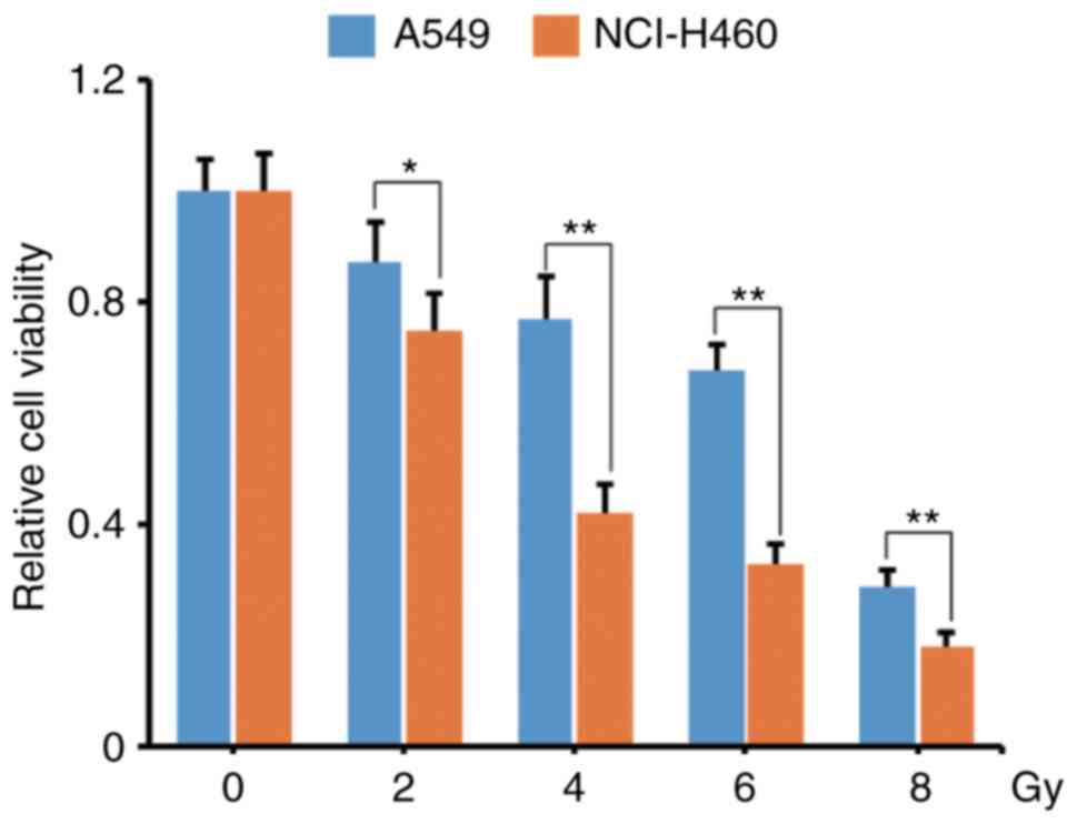
Differential radiosensitivity between
A549 and NCI-H460 cells
We used 2/4/6/8 Gy X-ray to irradiate A549 and
NCI-460 cells and compared the response of these two cell lines. As
revealed in Fig. 1, compared with
the unirradiated group, the relative cell viability of A549 vs.
NCI-H460 in 2/4/6/8 Gy IR groups were 87 vs. 75%, 77 vs. 42%, 68
vs. 33%, 29 vs. 18% (P<0.05), respectively. Notably, NCI-H460
cells were more radiosensitive than A549 cells, especially in the
4/6 Gy IR groups.
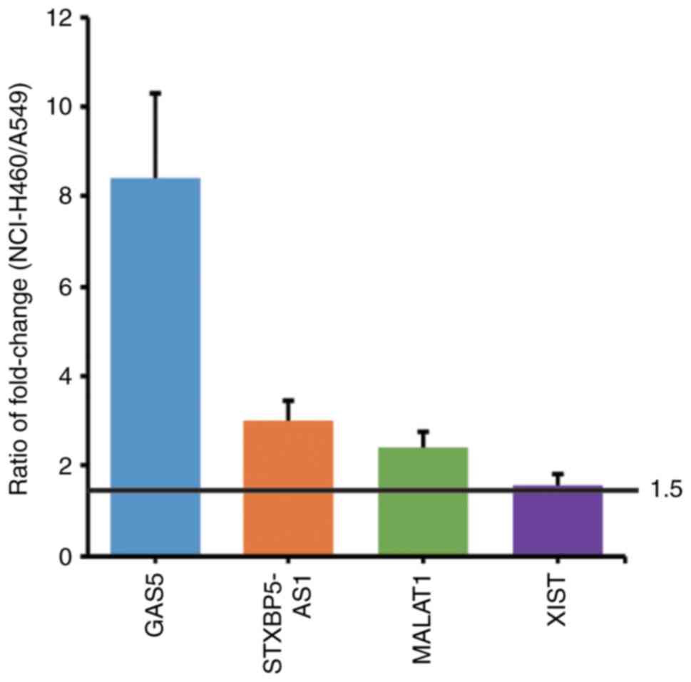
Screening of differentially expressed
lncRNAs between A549 and NCI-460 cells after IR
Differentially expressed lncRNAs of the irradiated
A549 and NCI-H460 cells were screened by RNA-Seq and analyzed. The
following standards were set for the selection of lncRNA
candidates: i) a FPKM fold-change (IR group/control group) of
NCI-H460 cells >2; ii) a ratio of FPKM fold-change (FPKM
fold-change of NCI-H460 cells/FPKM fold-change of A549 cells)
>1.5; iii) having definite biological functions. Finally, four
lncRNAs were identified: GAS5, STXBP5-AS1, MALAT1 and XIST. Their
FPKM values are listed in Table I.
The fold-change of each lncRNA and the ratios of fold-change
between the two cell lines are presented and compared in Fig. 2.
 | Table I.FPKM values of lncRNA GAS5,
STXBP5-AS1, MALAT1 and XIST. |
Table I.
FPKM values of lncRNA GAS5,
STXBP5-AS1, MALAT1 and XIST.
|
| FPKM (mean,
n=3) |
| FPKM (mean,
n=3) |
|
|---|
|
|
|
|
|
|
|---|
| Gene | NCI-H460 (CT) | NCI-H460 (IR) | Fold-change | A549 (CT) | A549 (IR) | Fold-change |
|---|
| GAS5 | 19.1 | 256.3 | 13.4 | 34.7 | 55.2 | 1.6 |
| STXBP5-AS1 | 22.5 | 153.8 | 6.8 | 15.3 | 34.8 | 2.3 |
| MALAT1 | 253.4 | 1023.3 | 4.0 | 410.1 | 706.5 | 1.7 |
| XIST | 54.4 | 114.5 | 2.1 | 41.1 | 52.1 | 1.3 |
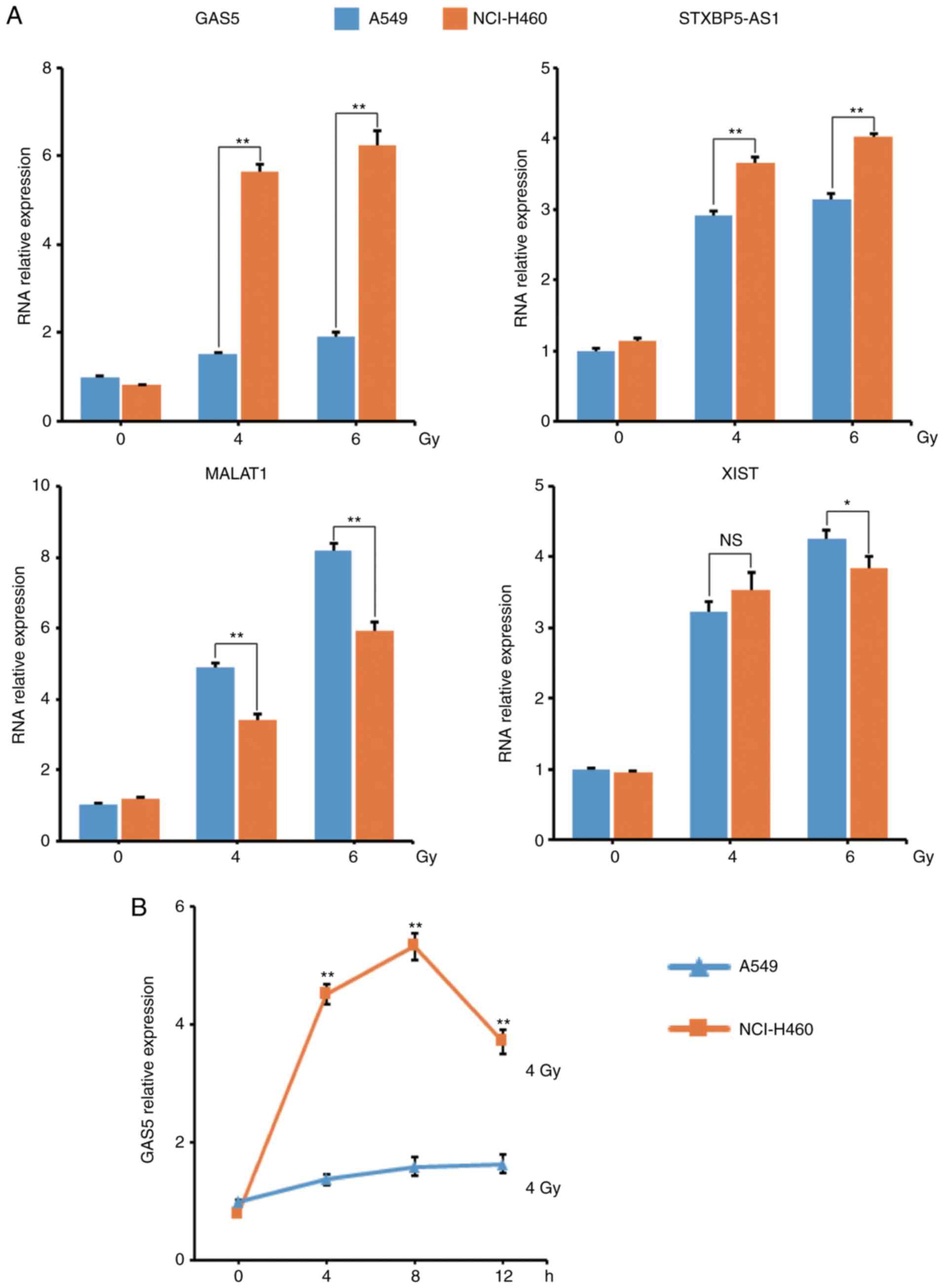
To verify the RNA-Seq result, the lncRNA levels in
these two cell lines were assessed using qPCR at 8 h post-IR. The
results revealed that lncRNA GAS5 had the most marked change and
the qPCR result was in line with the RNA-Seq result (Fig. 3A). The level of lncRNA GAS5 was then
monitored and it was revealed that the level of lncRNA GAS5 in
NCI-H460 cells was significantly higher than in A549 cells at
4/8/12 h post-4 Gy-IR (Fig.
3B).
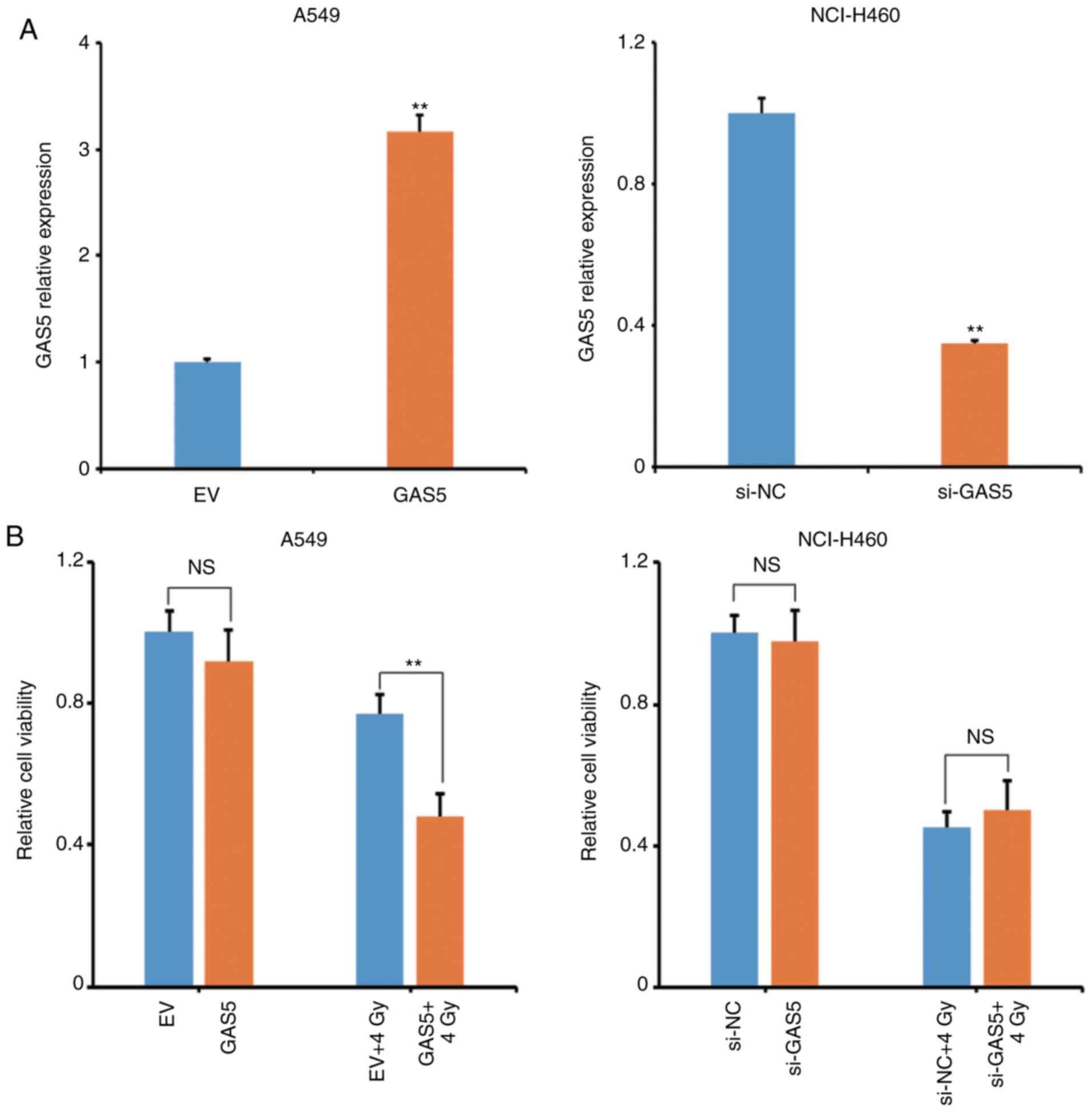
Overexpression of lncRNA GAS5
increases the radiosensitivity of A549 cells
To investigate the effect of lncRNA GAS5 on
IR-induced apoptosis, a GAS5-expressing plasmid was constructed and
transfected to A549 cells to observe whether it would enhance the
radiosensitivity. Concurrently, the lncRNA GAS5 siRNA was
transfected to NCI-H460 cells to observe whether it would weaken
the radiosensitivity. Fig. 4A
revealed that the transfection efficiency was satisfactory. The
level of lncRNA GAS5 in A549 cells was significantly increased and
in NCI-H460 cells it was significantly decreased. The viability of
transfectants at 12 h post-4 Gy-IR was then analyzed and it was
revealed that overexpression of lncRNA GAS5 significantly decreased
the viability of A549 cells (Fig.
4B, left panel). However, interfering of lncRNA GAS5 did not
affect the viability of NCI-H460 cells after IR (Fig. 4B, right panel). The apoptosis of
A549 transfectants was next analyzed and it was revealed that
overexpression of lncRNA GAS5 enhanced the IR-induced cell
apoptosis (Fig. 4C).
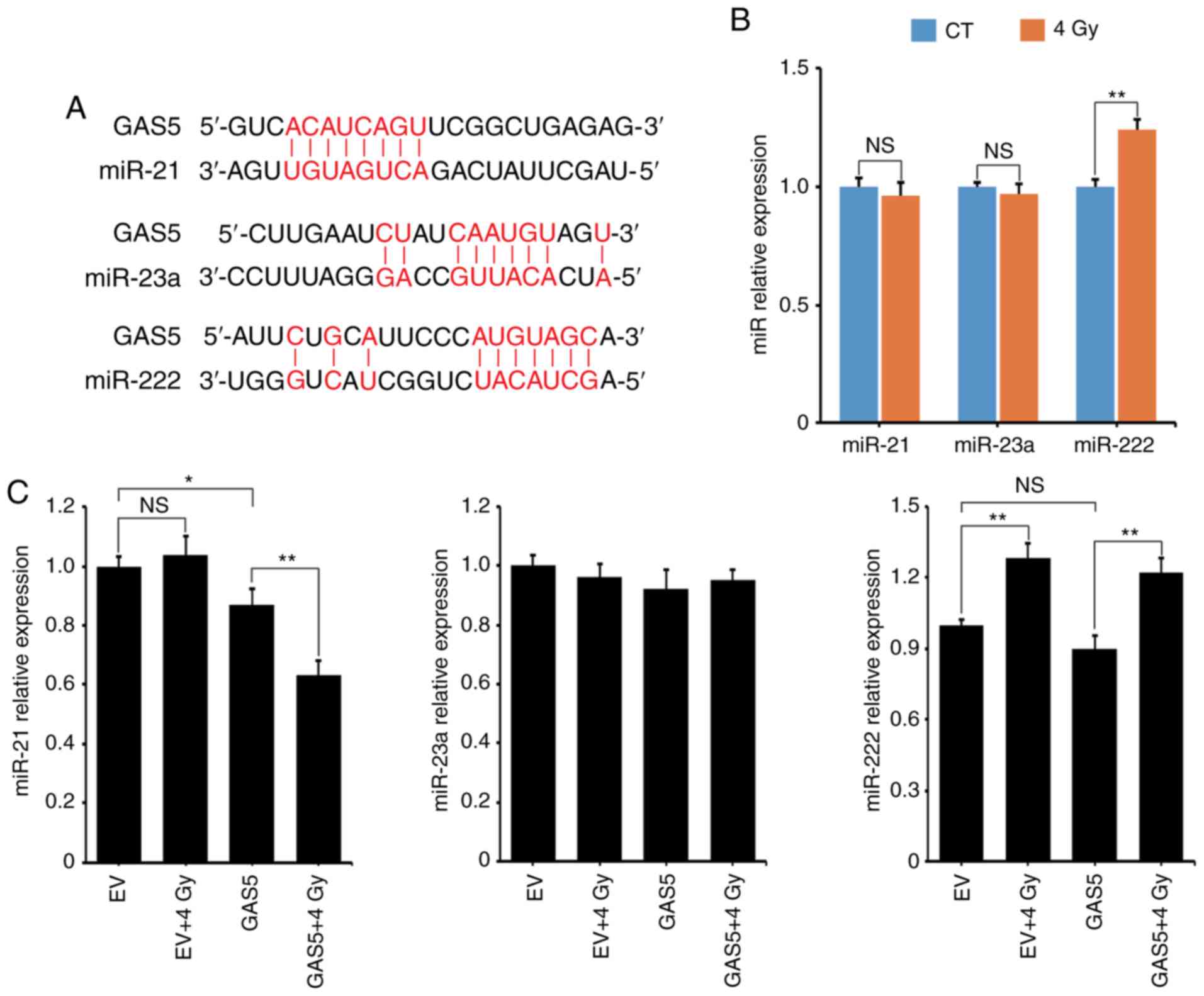
lncRNA GAS5 suppresses miR-21
expression during IR
It has been reported that lncRNA may serve as a
‘sponge’ to absorb miRNAs and regulate miRNA expression (24). In the present experiment, three
miRNAs that may interact with lncRNA GAS5 were selected: miR-21,
miR-23a and miR-222 (16,25,26).
According to the references, the binding sites of these three
miRNAs on lncRNA GAS5 are revealed in Fig. 5A. The expression levels of these
miRNAs at 8 h post-4 Gy-IR was assessed using qPCR and fit was
revealed that only miR-222 was upregulated (Fig. 5B). The expression levels of these
miRNAs in A549 transfectants was next assessed and it was revealed
that lncRNA GAS5 significantly suppressed the level of miR-21
during IR (Fig. 5C, left panel).
Although the level of miR-222 increased after IR, it was not
affected by the overexpression of lncRNA GAS5 (Fig. 5C, right panel). Additionally,
neither IR nor lncRNA GAS5 affected the level of miR-23a (Fig. 5C, middle panel).
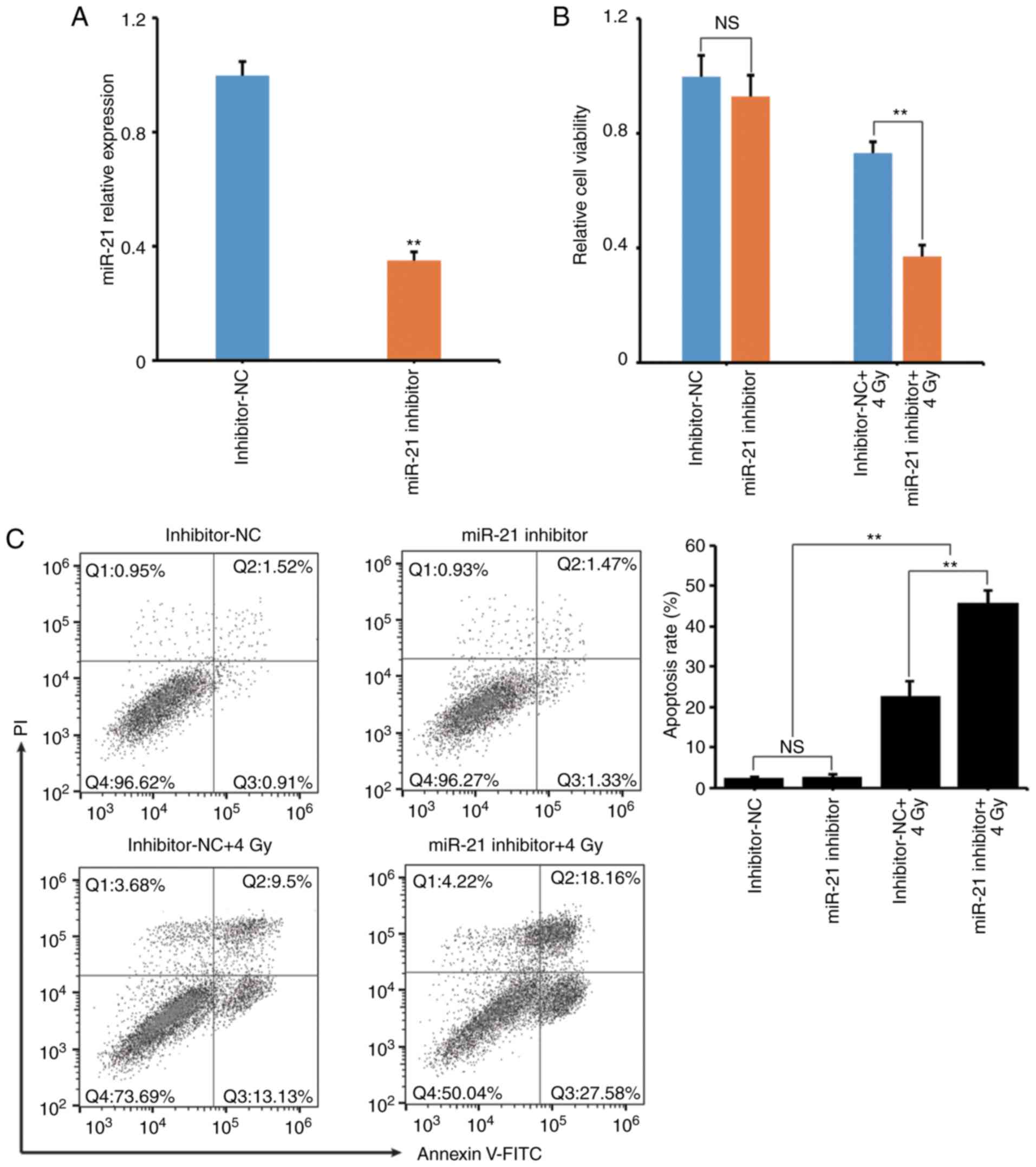
Inhibition of miR-21 increases the
radiosensitivity of A549 cells
To clarify the role of miR-21 in lncRNA GAS5-induced
cell apoptosis during IR, miR-21 expression was suppressed using a
miR-21 inhibitor. Fig. 6A reveals
that the transfection efficiency of the miR-21 inhibitor was
confirmed to be successful and the endogenous miR-21 expression was
significantly decreased. It was also revealed that suppression of
miR-21 significantly decreased the viability and increased the
IR-induced cell apoptosis rate (Fig. 6B
and C) of A549 cells.
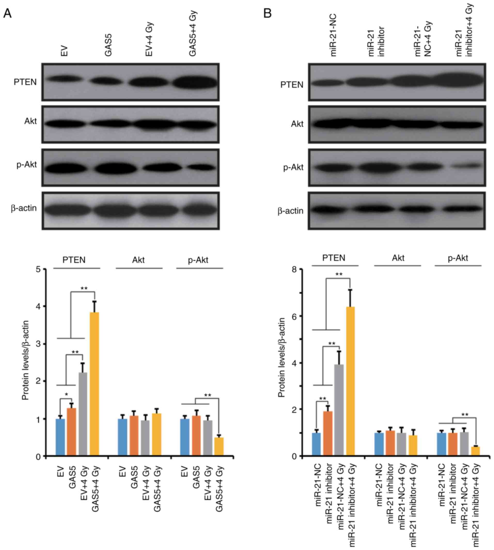
lncRNA GAS5 regulates the
radiosensitivity of A549 cells through the miR-21/PTEN/Akt
axis
PTEN is one of the most important target proteins of
miR-21, and the miR-21/PTEN/Akt axis has been revealed to play a
crucial role in various cellular activities (27–30).
In this experiment, the activation of the PTEN/Akt pathway was also
assessed in our experimental model. In Fig. 7, western blot results revealed that
overexpression of lncRNA GAS5 and suppression of miR-21 upregulated
the expression level of PTEN but did not affect the phosphorylation
of Akt in A549 cells. However, 4 Gy-IR significantly stimulated the
expression of PTEN and suppressed the phosphorylation of Akt.
Notably, overexpression of lncRNA GAS5 and inhibition of miR-21
exacerbated the IR-induced upregulation of PTEN and the
phosphorylation of Akt in A549 cells. These data indicated that
lncRNA GAS5 may function as a radiosensitizer through the
regulation of the miR-21/PTEN/Akt axis.
Discussion
LncRNA GAS5 was originally identified from a serum
starvation-induced cell growth arrest model (31). It was revealed to accumulate in
cells and sensitize cells to apoptosis by suppressing the
glucocorticoid-mediated induction of several responsive genes
(32). In recent studies, lncRNA
GAS5 has been revealed to be implicated in tumorigenesis and tumor
development in a variety of cancers (33–36).
However, the role of lncRNA GAS5 in NSCLC radiotherapy is still not
fully investigated.
In the present study, it was revealed that NSCLC
NCI-H460 cells were more prone to apoptosis than A549 cells during
IR. The correlation between radiosensitivity and the unique lncRNAs
was investigated. Using RNA-Seq, it was demonstrated that in
NCI-H460 cells four well-characterized lncRNAs, GAS5, STXBP5-AS1,
MALAT1 and XIST, were significantly upregulated after IR, among
which lncRNA GAS5 was the most differentially expressed. It was
also demonstrated that overexpression of lncRNA GAS5 could enhance
apoptosis of A549 cells. However, knockdown of lncRNA GAS5 did not
affect the radiosensitivity of NCI-H460 cells. These data indicated
that lncRNA GAS5 was sufficient but not necessary for the
radiosensitivity of NSCLC cells.
One important way that lncRNAs exert their
regulatory function is to serve as competing endogenous RNAs to
negatively regulate the biological activity of miRNAs. A number of
studies have reported the interaction between lncRNAs and miRNAs in
various physiological and pathological processes. For example, Lai
et al demonstrated that downregulation of lncRNA CCAT1
improved the radiosensitivity of breast cancer cells by negatively
regulating miR-148b (11). Wu et
al demonstrated that knockdown of lncRNA PVT1 improved the
radiosensitivity of NSCLC cells by directly interacting with
miR-195 (12). In the present
study, the expression of three miRNAs that may be targets of lncRNA
GAS5 were assessed and it was demonstrated that only miR-21 was
affected by lncRNA GAS5 inirradiated A549 cells. The direct
interaction between lncRNA GAS5 and miR-21 has been revealed by
several research groups using a luciferase assay (37,38).
The present results indicated that although overexpression of
lncRNA GAS5 could downregulate the level of miR-21 under normal
conditions, this suppressive effect was markedly increased during
IR. These data indicated that the interaction between lncRNA GAS5
and miR-21 may be an IR-specific response.
PTEN, a tumor suppressor, plays a critical role in
ceRNA networks (39). PTEN mRNA is
the competing target of many miRNAs, e.g. miR-624-3p, miR-130a, and
miR-21 (40–42). There are several potential binding
sites of miR-21 on the PTEN 3′UTR, such as GAUAAG and UCUG*UG*GCUA
(43,44). PTEN is an inhibitor of the PI3K/Akt
signaling pathway (45), and the
PTEN/Akt axis is a pivotal regulatory pathway in IR-induced cell
apoptosis. He et al demonstrated that overexpression of
circVRK1 could reverse the radioresistance of esophageal squamous
carcinoma cells by regulating the miR-624-3p/PTEN/PI3K/Akt
signaling pathway (40). Wang et
al reported that miR-29a regulated the radiosensitivity of
human intestinal cells by activating the PTEN/PI3K/Akt signaling
pathway (46). The present study
indicated that overexpression of lncRNA GAS5 or suppression of
miR-21 could increase the level of PTEN and decrease the
phosphorylation of Akt, especially during the IR process. This
indicated that lncRNA GAS5 enhanced the radiosensitivity of A549
cells by regulating the miR-21/PTEN/Akt pathway.
In conclusion, the present study revealed that
lncRNA GAS5 was associated with the distinct radiosensitivity of
NSCLC. Overexpression of lncRNA GAS5 could increase the IR-induced
cell apoptosis of A549 cells. Therefore, lncRNA GAS5 has a great
potential of being used as a radiosensitizer or a radiosensitivity
indicator in clinic radiotherapy. However, to draw a more solid
conclusion, additional cell lines, in vivo animal
experiments and clinical investigation should be carried out in
future work.
Acknowledgements
Not applicable.
Funding
The present study was supported by grants from the
Health Special Fund from Jilin Province Department of Finance no.
2018SCZWSZX-048 to XS), the Jilin International Collaboration Grant
no. 20180414065GH to DY) and the Norman Bethune Program of Jilin
University (Bethune plan to DY).
Availability of data and materials
The datasets used and analyzed during the current
study are available from the corresponding author on reasonable
request.
Authors' contributions
XS and BG contributed to the conception and design
of the study. XS and DY drafted the study. LC, PR and YZ
contributed to the acquisition, analysis and interpretation of
data. DY also revised this study critically for important
intellectual content. All the authors have read and approve the
final article to be submitted and agree to be accountable for all
aspects of the research in ensuring that the accuracy or integrity
of any part of the work are appropriately investigated and
resolved.
Ethics approval and consent to
participate
Not applicable.
Patient consent for publication
Not applicable.
Competing interests
The authors declare that they have no competing
interests.
References
|
1
|
Bray F, Ferlay J, Soerjomataram I, Siegel
RL, Torre LA and Jemal A: Global cancer statistics 2018: GLOBOCAN
estimates of incidence and mortality worldwide for 36 cancers in
185 countries. CA Cancer J Clin. 68:394–424. 2018. View Article : Google Scholar : PubMed/NCBI
|
|
2
|
Theys J, Yahyanejad S, Habets R, Span P,
Dubois L, Paesmans K, Kattenbeld B, Cleutjens J, Groot AJ,
Schuurbiers OCJ, et al: High NOTCH activity induces radiation
resistance in non small cell lung cancer. Radiother Oncol.
108:440–445. 2013. View Article : Google Scholar : PubMed/NCBI
|
|
3
|
Baker S, Dahele M, Lagerwaard FJ and Senan
S: A critical review of recent developments in radiotherapy for
non-small cell lung cancer. Radiat Oncol. 11:1152016. View Article : Google Scholar : PubMed/NCBI
|
|
4
|
Djebali S, Davis CA, Merkel A, Dobin A,
Lassmann T, Mortazavi A, Tanzer A, Lagarde J, Lin W, Schlesinger F,
et al: Landscape of transcription in human cells. Nature.
489:101–108. 2012. View Article : Google Scholar : PubMed/NCBI
|
|
5
|
Hsiao KY, Sun HS and Tsai SJ: Circular
RNA-new member of noncoding RNA with novel functions. Exp Biol Med
(Maywood). 242:1136–1141. 2017. View Article : Google Scholar : PubMed/NCBI
|
|
6
|
Barbarotto E, Schmittgen TD and Calin GA:
MicroRNAs and cancer: Profile, profile, profile. Int J Cancer.
122:969–977. 2008. View Article : Google Scholar : PubMed/NCBI
|
|
7
|
Mercer TR, Dinger ME and Mattick JS: Long
non-coding RNAs: Insights into functions. Nat Rev Genet.
10:155–159. 2009. View
Article : Google Scholar : PubMed/NCBI
|
|
8
|
Wright MW and Bruford EA: Naming ‘junk’:
Human non-protein coding RNA (ncRNA) gene nomenclature. Hum
Genomics. 5:90–98. 2011. View Article : Google Scholar : PubMed/NCBI
|
|
9
|
Ponting CP, Oliver PL and Reik W:
Evolution and functions of long noncoding RNAs. Cell. 136:629–641.
2009. View Article : Google Scholar : PubMed/NCBI
|
|
10
|
Wei S, Du M, Jiang Z, Hausman GJ, Zhang L
and Dodson MV: Long noncoding RNAs in regulating adipogenesis: New
RNAs shed lights on obesity. Cell Mol Life Sci. 73:2079–2087. 2016.
View Article : Google Scholar : PubMed/NCBI
|
|
11
|
Lai Y, Chen Y, Lin Y and Ye L:
Down-regulation of LncRNA CCAT1 enhances radiosensitivity via
regulating miR-148b in breast cancer. Cell Biol Int. 42:227–236.
2018. View Article : Google Scholar : PubMed/NCBI
|
|
12
|
Wu D, Li Y, Zhang H and Hu X: Knockdown of
Lncrna PVT1 enhances radiosensitivity in non-small cell lung cancer
by sponging mir-195. Cell Physiol Biochem. 42:2453–2466. 2017.
View Article : Google Scholar : PubMed/NCBI
|
|
13
|
Ricciuti B, Mencaroni C, Paglialunga L,
Paciullo F, Crinò L, Chiari R and Metro G: Long noncoding RNAs: New
insights into non-small cell lung cancer biology, diagnosis and
therapy. Med Oncol. 33:182016. View Article : Google Scholar : PubMed/NCBI
|
|
14
|
Sun M, Jin FY, Xia R, Kong R, Li JH, Xu
TP, Liu YW, Zhang EB, Liu XH and De W: Decreased expression of long
noncoding RNA GAS5 indicates a poor prognosis and promotes cell
proliferation in gastric cancer. BMC Cancer. 14:3192014. View Article : Google Scholar : PubMed/NCBI
|
|
15
|
Li W, Huang K, Wen F, Cui G, Guo H and
Zhao S: Genetic variation of lncRNA GAS5 contributes to the
development of lung cancer. Oncotarget. 8:91025–91029.
2017.PubMed/NCBI
|
|
16
|
Cao L, Chen J, Ou B, Liu C, Zou Y and Chen
Q: GAS5 knockdown reduces the chemo-sensitivity of non-small cell
lung cancer (NSCLC) cell to cisplatin (DDP) through regulating
miR-21/PTEN axis. Biomed Pharmacother. 93:570–579. 2017. View Article : Google Scholar : PubMed/NCBI
|
|
17
|
Xue Y, Ni T, Jiang Y and Li Y: Long
noncoding RNA GAS5 inhibits tumorigenesis and enhances
radiosensitivity by suppressing miR-135b expression in non-small
cell lung cancer. Oncol Res. 25:1305–1316. 2017. View Article : Google Scholar : PubMed/NCBI
|
|
18
|
Cron KR, Zhu K, Kushwaha DS, Hsieh G,
Merzon D, Rameseder J, Chen CC, D'Andrea AD and Kozono D:
Proteasome inhibitors block DNA repair and radiosensitize non-small
cell lung cancer. PLoS One. 8:e737102013. View Article : Google Scholar : PubMed/NCBI
|
|
19
|
Kim W, Youn H, Seong KM, Yang HJ, Yun YJ,
Kwon T, Kim YH, Lee JY, Jin YW and Youn B: PIM1-activated PRAS40
regulates radioresistance in non-small cell lung cancer cells
through interplay with FOXO3a, 14-3-3 and protein phosphatases.
Radiat Res. 176:539–552. 2011. View
Article : Google Scholar : PubMed/NCBI
|
|
20
|
Liu L, Wang HJ, Meng T, Lei C, Yang XH,
Wang QS, Jin B and Zhu JF: lncRNA GAS5 inhibits cell migration and
invasion and promotes autophagy by targeting miR-222-3p via the
GAS5/PTEN-signaling pathway in CRC. Mol Ther Nucleic Acids.
17:644–656. 2019. View Article : Google Scholar : PubMed/NCBI
|
|
21
|
Huang J, Xie N, Huang H, Yao J and Hu W:
Long noncoding RNA STXBP5-AS1 inhibits cell proliferation,
migration, and invasion via preventing the PI3K/AKT against STXBP5
expression in non-small-cell lung carcinoma. J Cell Biochem.
18:280232018.
|
|
22
|
Zhang YL, Li XB, Hou YX, Fang NZ, You JC
and Zhou QH: The lncRNA XIST exhibits oncogenic properties via
regulation of miR-449a and Bcl-2 in human non-small cell lung
cancer. Acta Pharmacol Sin. 38:371–381. 2017. View Article : Google Scholar : PubMed/NCBI
|
|
23
|
Livak KJ and Schmittgen TD: Analysis of
relative gene expression data using real-time quantitative PCR and
the 2(-Delta Delta C(T)) method. Methods. 25:402–408. 2001.
View Article : Google Scholar : PubMed/NCBI
|
|
24
|
Wei Y, Yan Z, Wu C, Zhang Q, Zhu Y, Li K
and Xu Y: Integrated analysis of dosage effect lncRNAs in lung
adenocarcinoma based on comprehensive network. Oncotarget.
8:71430–71446. 2017. View Article : Google Scholar : PubMed/NCBI
|
|
25
|
Dong Z, Li S, Wang X, Si L, Ma R, Bao L
and Bo A: lncRNA GAS5 restrains CCl4-induced hepatic fibrosis by
targeting miR-23a through the PTEN/PI3K/Akt signaling pathway. Am J
Physiol Gastrointest Liver Physiol. 316:G539–G550. 2019. View Article : Google Scholar : PubMed/NCBI
|
|
26
|
Li Y, Gu J and Lu H: The GAS5/miR-222 axis
regulates proliferation of gastric cancer cells through the
PTEN/Akt/mTOR pathway. Dig Dis Sci. 62:3426–3437. 2017. View Article : Google Scholar : PubMed/NCBI
|
|
27
|
Wickramasinghe NS, Manavalan TT, Dougherty
SM, Riggs KA, Li Y and Klinge CM: Estradiol downregulates miR-21
expression and increases miR-21 target gene expression in MCF-7
breast cancer cells. Nucleic Acids Res. 37:2584–2595. 2009.
View Article : Google Scholar : PubMed/NCBI
|
|
28
|
Liu HY, Zhang YY, Zhu BL, Feng FZ, Yan H,
Zhang HY and Zhou B: MiR-21 regulates the proliferation and
apoptosis of ovarian cancer cells through PTEN/PI3K/AKT. Eur Rev
Med Pharm Sci. 23:4149–4155. 2019.
|
|
29
|
Liu H, Wang J, Tao Y, Li X, Qin J, Bai Z,
Chi B, Yan W and Chen X: Curcumol inhibits colorectal cancer
proliferation by targeting miR-21 and modulated PTEN/PI3K/Akt
pathways. Life Sci. 221:354–361. 2019. View Article : Google Scholar : PubMed/NCBI
|
|
30
|
Zhu Y, Tang H, Zhang L, Gong L, Wu G, Ni J
and Tang X: Suppression of miR-21-3p enhances TRAIL-mediated
apoptosis in liver cancer stem cells by suppressing the
PI3K/Akt/Bad cascade via regulating PTEN. Cancer Manag Res.
11:955–968. 2019. View Article : Google Scholar : PubMed/NCBI
|
|
31
|
Schneider C, King RM and Philipson L:
Genes specifically expressed at growth arrest of mammalian cells.
Cell. 54:787–793. 1988. View Article : Google Scholar : PubMed/NCBI
|
|
32
|
Kino T, Hurt DE, Ichijo T, Nader N and
Chrousos GP: Noncoding RNA gas5 is a growth arrest- and
starvation-associated repressor of the glucocorticoid receptor. Sci
Signal. 3:ra82010. View Article : Google Scholar : PubMed/NCBI
|
|
33
|
Mei Y, Si J, Wang Y, Huang Z, Zhu H, Feng
S, Wu X and Wu L: Long noncoding RNA GAS5 suppresses tumorigenesis
by inhibiting miR-23a expression in non-small cell lung cancer.
Oncol Res. 25:1027–1037. 2017. View Article : Google Scholar : PubMed/NCBI
|
|
34
|
Bian D, Shi W, Shao Y, Li P and Song G:
Long non-coding RNA GAS5 inhibits tumorigenesis via miR-137 in
melanoma. Am J Transl Res. 9:1509–1520. 2017.PubMed/NCBI
|
|
35
|
Guo LJ, Zhang S, Gao B, Jiang Y, Zhang XH,
Tian WG, Hao S, Zhao JJ, Zhang G, Hu CY, et al: Low expression of
long non-coding RNA GAS5 is associated with poor prognosis of
patients with thyroid cancer. Exp Mol Pathol. 102:500–504. 2017.
View Article : Google Scholar : PubMed/NCBI
|
|
36
|
Yan H, Zhang DY, Li X, Yuan XQ, Yang YL,
Zhu KW, Zeng H, Li XL, Cao S, Zhou HH, et al: Long non-coding RNA
GAS5 polymorphism predicts a poor prognosis of acute myeloid
leukemia in Chinese patients via affecting hematopoietic
reconstitution. Leuk Lymphoma. 58:1948–1957. 2017. View Article : Google Scholar : PubMed/NCBI
|
|
37
|
Wen Q, Liu Y, Lyu H, Xu X, Wu Q, Liu N,
Yin Q, Li J and Sheng X: Long noncoding RNA GAS5, which acts as a
tumor suppressor via microRNA 21, regulates cisplatin resistance
expression in cervical cancer. Int J Gynecol Cancer. 27:1096–1108.
2017. View Article : Google Scholar : PubMed/NCBI
|
|
38
|
Ma N, Li S, Zhang Q, Wang H, Qin H and
Wang S: Long non-coding RNA GAS5 inhibits ovarian cancer cell
proliferation via the control of microRNA-21 and SPRY2 expression.
Exp Ther Med. 16:73–82. 2018.PubMed/NCBI
|
|
39
|
Poliseno L and Pandolfi PP: PTEN ceRNA
networks in human cancer. Methods. 77-78:41–50. 2015. View Article : Google Scholar : PubMed/NCBI
|
|
40
|
He Y, Mingyan E, Wang C, Liu G, Shi M and
Liu S: CircVRK1 regulates tumor progression and radioresistance in
esophageal squamous cell carcinoma by regulating
miR-624-3p/PTEN/PI3K/AKT signaling pathway. Int J Biol Macromol.
125:116–123. 2019. View Article : Google Scholar : PubMed/NCBI
|
|
41
|
Wei H, Cui R, Bahr J, Zanesi N, Luo Z,
Meng W, Liang G and Croce CM: Mir-130a deregulates PTEN and
stimulates tumor growth. Cancer Res. 77:6168–6178. 2017. View Article : Google Scholar : PubMed/NCBI
|
|
42
|
Wu Y, Song Y, Xiong Y, Wang X, Xu K, Han
B, Bai Y, Li L, Zhang Y and Zhou L: MicroRNA-21 (Mir-21) promotes
cell growth and invasion by repressing tumor suppressor PTEN in
colorectal cancer. Cell Physiol Biochem. 43:945–958. 2017.
View Article : Google Scholar : PubMed/NCBI
|
|
43
|
He C, Dong X, Zhai B, Jiang X, Dong D, Li
B, Jiang H, Xu S and Sun X: MiR-21 mediates sorafenib resistance of
hepatocellular carcinoma cells by inhibiting autophagy via the
PTEN/Akt pathway. Oncotarget. 6:28867–28881. 2015. View Article : Google Scholar : PubMed/NCBI
|
|
44
|
Liu H, Li H, Jin L, Li G, Hu S, Ning C,
Guo J, Shuai S, Li X and Li M: Long noncoding RNA GAS5 suppresses
3T3-L1 cells adipogenesis through miR-21a-5p/PTEN signal pathway.
DNA Cell Biol. 37:767–777. 2018. View Article : Google Scholar : PubMed/NCBI
|
|
45
|
Haddadi N, Lin Y, Travis G, Simpson AM,
Nassif NT and McGowan EM: PTEN/PTENP1: ‘Regulating the regulator of
RTK-dependent PI3K/Akt signalling’, new targets for cancer therapy.
Mol Cancer. 17:372018. View Article : Google Scholar : PubMed/NCBI
|
|
46
|
Wang J, Xu J, Fu J, Yuan D, Guo F, Zhou C
and Shao C: MiR-29a regulates radiosensitivity in human intestinal
cells by targeting PTEN gene. Radiat Res. 186:292–301. 2016.
View Article : Google Scholar : PubMed/NCBI
|