Introduction
Osteosarcoma is a common malignant bone tumor, which
is highly lethal to adolescents and the elderly (1,2).
Osteosarcoma cells have strong metastatic and invasion abilities,
the disease could be aggravated by lung metastases at early stages
(3). Although numerous studies in
recent years have provided diverse treatment options, including
gene therapy targeting long non-coding RNA (lncRNA) or microRNA
(miR/miRNA) for osteosarcoma, the disease is still not fully
understood (4,5).
Long non-coding RNA (lncRNA) is a type of the
non-coding RNA molecule composed of >200 nucleotides (6,7). A
study showed that lncRNAs play a critical regulatory role in the
proliferation, differentiation and apoptosis of osteosarcoma cells
(8). Several studies also suggested
that lncRNA expression could regulate the proliferation, migration
and invasion of multiple types of malignant tumor cells (9,10).
LncRNA prostate cancer-associated transcript 6 (lncRNA PCAT6)
promoted the proliferation of prostate cancer cells in an
androgen-independent manner (11).
Furthermore, high levels of lncRNA PCAT6 also promoted the
proliferation, migration and invasion of non-small cell lung cancer
cells and ovarian cancer (12,13).
LncRNA PCAT6 was reported to act as a miRNA sponge to regulate the
proliferation, migration and invasion of osteosarcoma (14). However, the effect of lncRNA PCAT6
on the occurrence of osteosarcoma remains to be elucidated.
P53 is a classic tumor suppressor gene (15). Higher levels of P53 expression could
inhibit the proliferation and metastasis of breast, liver and lung
cancer cells (15). P21, also known
as CDKN1A, plays a crucial role in cell cycle progression (16). P21 could also suppress the
proliferation of multiple cancer cells by inducing cell cycle
arrest (17). A study revealed that
P53 could activate the expression of P21 and therefore enhance the
antitumor effects of P53/P21 (18).
Another study indicated that MDM2 could promote the occurrence and
development of malignant tumors by inducing P53 ubiquitination
(19). Several studies also
indicated that E3 ubiquitin-protein ligase Mdm2 (MDM2), which
negatively regulates p53 protein, could promote the development of
ovarian (20) and lung cancer
(21). However, whether lncRNA
PCAT6 could promote the development of osteosarcoma by promoting
the expression of MDM2 is unclear.
The present study established lncRNA PCAT6-
overexpressing osteosarcoma cell lines. Subsequently, the
proliferation, migration and invasion of these cells were
determined with Cell Counting Kit (CCK)-8, wound healing and
Transwell assays, respectively. MDM2 expression was then suppressed
in the lncRNA PCAT6-overexpressing osteosarcoma cell line by
transfection. Following which, the proliferation and metastasis of
these cells were detected again. The results of these assays could
reveal the effect of lncRNA PCAT6 on osteosarcoma development.
Materials and methods
Cell culture and transfection
Normal osteoblasts (hFOB1.19) and human osteosarcoma
cell lines (Saos2, MG63, U2OS and HOS) used in the present study
were purchased from American Type Culture Collection. Cells were
cultured with RPMI-1640 medium (HyClone; Cytiva) supplemented with
10% FBS and 1% penicillin/streptomycin (Gibco; Thermo Fisher
Scientific, Inc.). Cells were cultured in 37°C humid atmosphere
with 5% CO2. LncRNA PCAT6 overexpression lentiviruses (4
µg/ml; 1 ml) were designed and purchased from Shanghai GeneChem
Co., Ltd. 293T cells (American Type Culture Collection) were used
to package lncRNA PCAT6 overexpression lentivirus vectors or empty
lentivirus vectors (NC groups). The MG63 cells in the control group
were not treated with lentiviruses and transfection reagent. Small
interfering RNA (si)-MDM2 (si-MDM2-1, 5′CAGCCATCAACTTCTAGTA3′;
si-MDM2-2, 5′CCACCTCACAGATTCCAGCTTCAAGAGAGCTGGAATCTGTGAGGTGG-3′)
lentivirus (4 µg/ml; 1 ml) and corresponding vector were also
obtained from Shanghai GeneChem Co., Ltd. Polybrene (Shanghai
GeneChem Co., Ltd.) was used to increase transfection efficacy.
After 24 h of transfection, MG63 cells were used for further
experiments.
Animal assays
A total of 30 BALB/c nude mice (male; 4-week-old;
18–20 g) were obtained from the Shanghai Lingchang Biotechnology
Company (Shanghai, China). Mice were raised in a sterile laminar
flow cabinet with a 12-h light/dark cycle at 25±1°C and 40–60%
humidity and free access to food and water.
Subsequently, MG63 cells (1×10⁶) were
injected into the thigh joint of nude mice by subcutaneous
injection. After two weeks, these mice were anesthetized with
ketamine (80 mg/kg) and xylazine (7 mg/kg). The mice were then
placed in a carbon dioxide (CO2) euthanasia chamber
(Shanghai Yuyan Instruments Co., Ltd.) and sacrificed by excess
CO2. The controlled flow rate of CO2 was 20%
of the volume of the euthanasia chamber per minute. Once the animal
lost consciousness, the flow rate was increased to 100% of the
euthanasia chamber volume per minute. Mice tumors were collected
for subsequent experimentation. The percentage of weight loss was
also calculated using the following formula: (Daily weight-weight
on the first day)/weight on the first day ×100%. The weight and
tumor volume of mice were recorded each day. Tumor weight was
detected with an electronic balance. The formula for calculating
tumor volume of mice is as follows: Tumor volume=0.5 × length ×
width2. Cells transfected with pc-DNA PCAT6 or si-MDM2
were injected into the back of nude mice (1×10⁶ cells).
The mice were divided into four groups (n=7 per group):
pcDNA-negative control (NC), pcDNA-PCAT6, pcDNA-PCAT6 + si-MDM2 and
pcDNA-PCAT6 + RG7112 (5 µm; cat. no. HY-10959; MedChemExpress).
Two mice died during the experiments and were not
used to perform the analysis of the result. The possible causes
were as follows: i) The rapidly growing tumor led to the restricted
movement of the mice's forelimbs, which affected feeding and led to
death; or ii) the influences of the experimental environment. All
animal experiments were performed in accordance with the animal
experimental guidelines set by the National Institutes of Health
Guide for the Care and Use of Laboratory Animals. The study was
approved by the Experimental Animal Ethical Committee of Zhejiang
Hospital (approval no. JUMC2019-019).
CCK-8 assay
MG63 cells were plated into a 96-well plate
(2×10³ cells/well). Cell proliferation abilities were
detected using a CCK-8 assay (Dojindo Molecular Technologies, Inc.)
and performed according to the manufacturer's instructions. After
24 h, 10 µl CCK-8 solution (Dojindo Molecular Technologies, Inc.)
was diluted in culture medium and added into the 96-well plates.
Subsequently, cells were incubated at 37°C for 1 h. The absorbance
of the cells at a wavelength of 450 nm was detected with a
spectrophotometer (Thermo Fisher Scientific, Inc.). The other two
plates were detected after 48 and 72 h of culture.
Wound healing assay
Prior to experimentation, cells of different groups
were plated into six-well plates (4×10⁵ cells/well).
Once adhered, cells were cultured in serum-free medium for 12 h. A
scratch was then created with a pipette tip. The width of the
scratch was photographed and recorded at 0 h (magnification, ×100).
Subsequently, the width of the scratch was photographed after 24 h
(magnification, ×100) with a light microscope (Olympus
Corporation). The migration rates were calculated with the
following formula: Migration rate=[(width at 0 h-width at 24
h)/width of 0 h] ×100%.
Transwell assay
Cells were cultured in serum-free medium for 12 h
before experimentation. Cells (1×10⁵/ml) were then
plated into Transwell chambers (Corning Inc.). Complete medium with
10% FBS was also added to the lower Transwell chamber. Following 24
h, the cells in the opposite basement membrane of the Transwell
chamber was fixed with 4% paraformaldehyde for 10 min and then
stained with crystal violet for 5 min at room temperature (Thermo
Fisher Scientific, Inc.). Stained cells were calculated under a
light microscope (magnification, ×100; Olympus Corporation).
Hematoxylin and eosin (HE)
staining
Tumor tissues were collected and used for
experiments after mice euthanasia. After tumor tissues were fixed
in 4% paraformaldehyde for 48 h at 4°C, tumor tissues were embedded
with paraffin and then cut into 5-µm sections. Slices were
subsequently stained with hematoxylin and eosin (Thermo Fisher
Scientific, Inc.) for 5 min at room temperature. Three slices were
observed and photographed under a light microscope (magnification,
×200; Olympus Corporation).
Immunohistochemistry
Tumor tissues were fixed in 4% paraformaldehyde for
48 h at 4°C. Subsequently, tumor tissues were dehydrated and cut
into 5-µm sections. An appropriate amount of normal goat serum
(Beyotime Institute of Biotechnology) was added to the slide tissue
for non-specific antigen blocking for 30 min at 37°C. Subsequently,
Ki-67 expression in tumor tissues was detected with Ki-67 antibody
(1:5,000; cat. no. ab15580; Abcam) according to the manufacturer's
instructions. Following which, goat anti-rabbit biotinylated
secondary antibody (1:40,000; cat. no. ab7089; Abcam) was added to
detect the primary antibody. Neutral balsam was used to mount the
sections. The staining results were observed under a light
microscope (magnification, ×200; Olympus Corporation).
Reverse transcription-quantitative PCR
(RT-qPCR)
Total RNA was extracted from cells using
TRIzol® reagent (Invitrogen; Thermo Fisher Scientific,
Inc.). RNA was reverse transcribed into cDNA using a reverse
transcription kit (cat. no. 4897030001; Roche Diagnostics)
according to the manufacturer's instructions. SYBR Green (Thermo
Fisher Scientific, Inc.) was used as the fluorescence indicator
during the cDNA amplification process. The following thermocycling
conditions were used for the qPCR: Initial denaturation at 95°C for
30 sec; followed by 40 cycles of denaturation at 95°C for 5 sec and
annealing and extension at 60°C for 30 sec. The expression of
targeted genes was calculated using the 2−∆∆Cq method
(22). The following primer pairs
were used for the qPCR: MDM2 forward, 5′-AGTAGCAGTGAATCTACAGGGA-3′
and reverse, 5′-CTGATCCAACCAATCACCTGAAT-3′; lncRNA PCAT6 forward,
5′-CCCCTCCTTACTCTTGGACAAC-3′ and reverse,
5′-GACCGAATGAGGATGGAGACAC-3′ and GAPDH forward,
5′-AGAAGGCTGGGGCTCATTTG-3′ and reverse,
5′-AGGGGCCATCCACAGTCTTC-3′.
Western blotting
Total protein (20 µg) was extracted from cells or
tumor tissues using RIPA buffer (Beyotime Institute of
Biotechnology). Protein concentration was determined using a BCA
assay. Proteins were separated via 12% SDS-PAGE (Beyotime Institute
of Biotechnology) and transferred to PVDF membranes (EMD
Millipore). Subsequently, membranes were blocked in 5% skim milk
powder solution at room temperature for 1 h and incubated with
primary antibodies overnight at 4°C. The primary antibodies used
were as follows: MMP-2 (1:1,000; cat. no. ab97779; Abcam), MMP-9
(1:1,000; cat. no. ab38898; Abcam), P21 (1:5,000; cat. no.
ab109520; Abcam), P53 cat. no. (1:5,000; ab26; Abcam), MDM2
(1:1,000; cat. no. ab16895; Abcam) and GAPDH (1:5,000; cat. no.
ab9485; Abcam). Subsequently, membranes were incubated with
HRP-conjugated secondary antibodies (1:10,000; cat. nos. ab97040
and ab97080; Abcam) at room temperature for 1 h. On the second day,
membranes were washed with PBS-Tween-20 (0.05%) three times.
Finally, the bands were visualized with chemiluminescence reagent
(EMD Millipore), followed by the analysis of gray value of protein
bands using ImageJ software (version 1.46; National Institutes of
Health).
Statistical analysis
Data were analyzed using GraphPad Prism 8.0
(GraphPad Software, Inc.). One-way ANOVA was used for comparison
among multiple groups, followed by Tukey's post hoc test. Data were
expressed as the mean ± SD. All experiments in were repeated three
times. P<0.05 was considered to indicate a statistically
significant difference.
Results
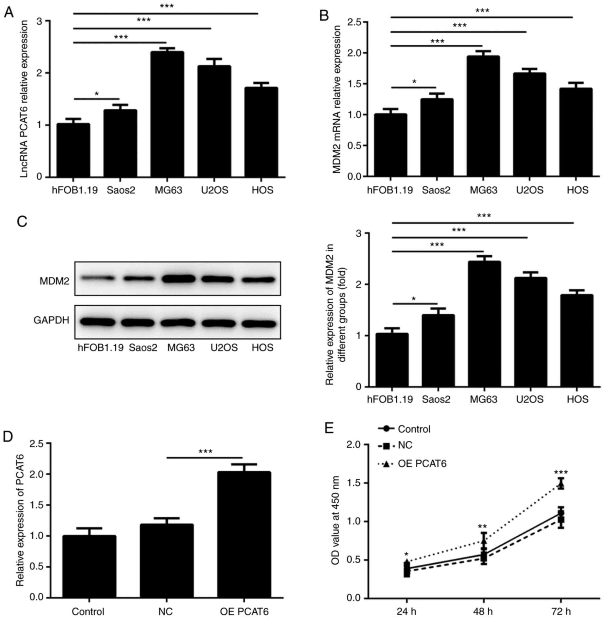
Overexpression of lncRNA PCAT6
promotes the proliferation of osteosarcoma cells
LncRNA PCAT6 was reported to be upregulated in
osteosarcoma tissue (14). The
present study detected the expression of lncRNA PCAT6 and MDM2 in
osteosarcoma cell lines (hFOB1.19, Saos2, MG63, U2OS and HOS). As
shown in Fig. 1A-C, lncRNA PCAT6
and MDM2 expression was highest in MG63 cells. Therefore, MG63
cells was selected for subsequent experiments. LncRNA
PCAT6-overexpressing osteosarcoma cells were then established.
Fig. 1D shows that lncRNA PCAT6
expression in the overexpression group was significantly higher
compared with the NC group. CCK-8 assays were performed to detect
the change in MG63 cell proliferation. Compared with the NC group,
the proliferation of MG63 cells significantly increased following
overexpression of lncRNA PCAT6 at 24, 48 and 72 h (Fig. 1E).
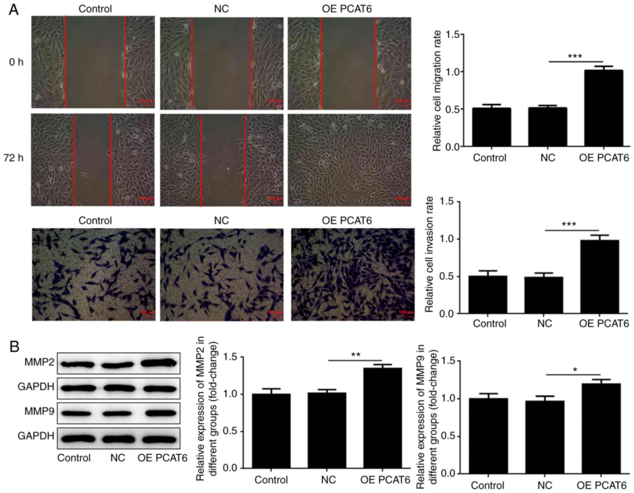
Overexpression of lncRNA PCAT6
enhances the migration and invasion of osteosarcoma cells
Wound healing and Transwell assays were performed to
detect the migration and invasion abilities of MG63 cells following
overexpression of lncRNA PCAT6. Based on the results (Fig. 2A), it was found that the migration
and invasion of MG63 cells significantly increased following
overexpression of lncRNA PCAT6 compared with the NC group.
Increased levels of MMP-2 and MMP-9 were closely correlated with
metastases of osteosarcoma (23,24).
Subsequently, MMP-2 and MMP-9 expression was detected via western
blotting. As shown in Fig. 2B,
MMP-2 and MMP-9 expression in MG63 cells significantly increased
following overexpression of lncRNA PCAT6 compared with the NC
group.
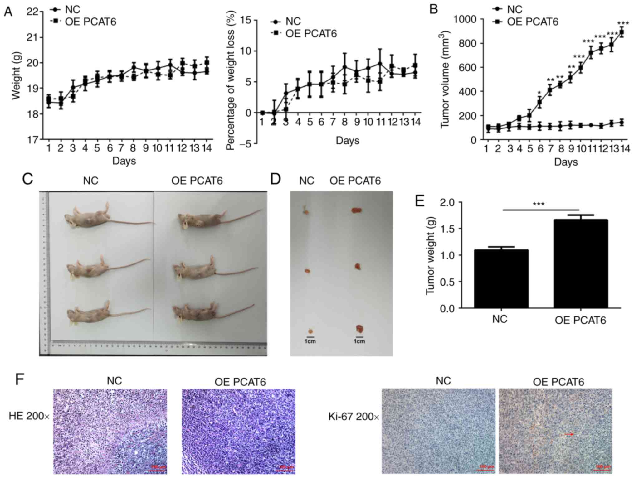
Overexpression of lncRNA PCAT6
promotes the proliferation of osteosarcoma cells in vivo
MG63 cells (negative control and overexpression
group) were injected into mice by subcutaneous injection. The
weight of these mice was measured each day for 14 days after
injection. There was no significant difference in the weight and
weight loss of mice between the PCAT6 overexpression group and
control group (Fig. 3A). The tumor
volume was significantly increased to varying degrees following
overexpression of lncRNA PCAT6 (Fig.
3B). The maximum tumor diameter measured was 0.94 cm. The tumor
weight significantly increased following overexpression of lncRNA
PCAT6 compared with the NC group (Fig.
3C-E). Additionally, the maximum percentage of tumor weight out
of total animal body weight was 8.59%. Tumor nuclei were heavily
stained and the atypia was obvious using HE staining in the OE
PCAT6 group compared with the NC group (Fig. 3F). Immunohistochemistry results
(Fig. 3F) also showed that ki-67
expression was enhanced in tumor tissues overexpressing lncRNA
PCAT6.
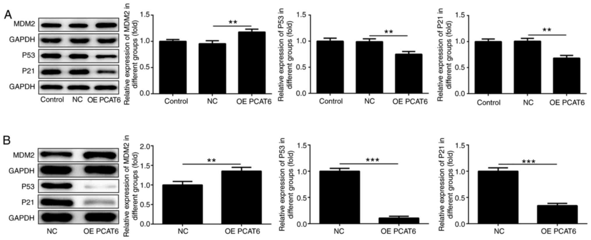
Overexpression of lncRNA PCAT6
promotes MDM2 expression
Subsequently, the expression of MDM2, P53 and P21 in
MG63 cells and tumors was detected. The results showed that MDM2
expression significantly increased following lncRNA PCAT6
overexpression compared with the NC group (Fig. 4A and B). However, the levels of P53
and P21 significantly decreased following overexpression of lncRNA
PCAT6 compared with the NC group.
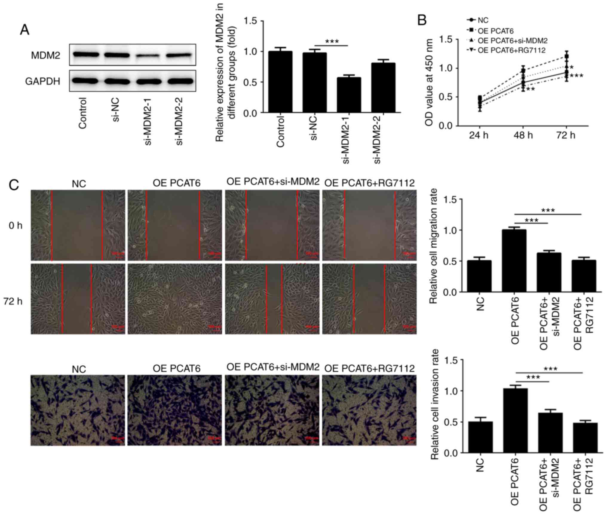
MDM2 knockdown weakens the
proliferation, migration and invasion of osteosarcoma cells
Lentiviruses were designed to establish
MDM2-knockdown osteosarcoma cells. As shown in Fig. 5A, MDM2 expression was inhibited
following transfection with si-MDM2. However, the inhibition
efficiency of si-MDM2-1 was higher compared with si-MDM2-2.
Therefore, si-MDM2-1 was selected for subsequent experiments. Next,
CCK-8 assays were performed to MG63 cell proliferation. The results
showed that MG63 cell proliferation was suppressed following MDM2
knockdown compared with the OE PCAT6 group at 48 and 72 h (Fig. 5B). Similarly, compared with the OE
PCAT6 group, the proliferation of these cells was also promoted
after treatment with RG7112, an MDM2 inhibitor. The migration and
invasion of MG63 cells were also significantly inhibited after MDM2
knockdown and RG7112 treatment compared with PCAT6 overexpression
(Fig. 5C).
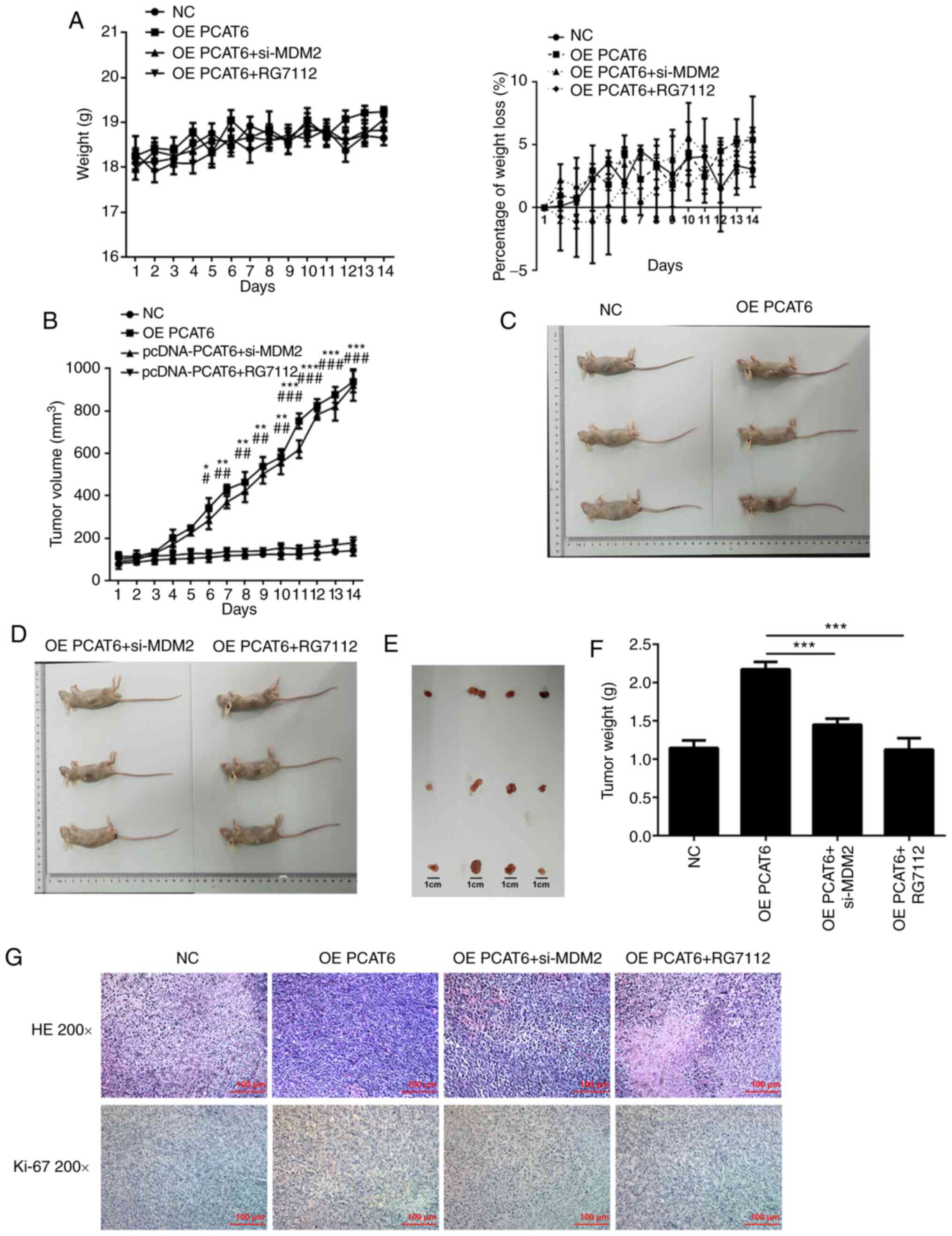
MDM2 knockdown suppresses the
proliferation of osteosarcoma cells in vivo
For these experiments, MG63 cells were injected into
nude mice by subcutaneous injection. There was no significant
difference in weight and weight loss between the mice of different
groups (Fig. 6A). The tumor volume
and tumor weight declined following MDM2 knockdown and RG7112
treatment (Fig. 6B and F). The mice
and tumors in different groups are shown in Fig. 6C-E. The maximum percentage of tumor
weight, out of total animal body weight, was 11.65% and the maximum
tumor diameter measured was 0.98 cm. Compared with the OE PCAT6
group, the nucleolus partially disappeared while the
nucleocytoplasmic ratio decreased in OE PCAT5+si-MDM2 and OE
PCAT5+RG7112 groups through HE staining and immunohistochemistry
results also showed that ki-67 expression was inhibited following
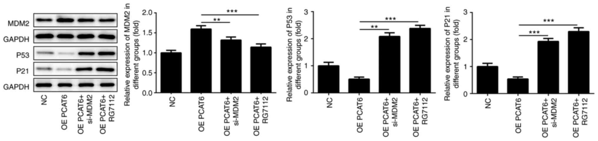
MDM2 knockdown and RG7112 treatment (Fig. 6G). In previous studies, we found
that lncRNA PCAT6 knockdown enhanced the expression of P53
(25). Therefore, the expression of
P53 and P21 after MDM2 knockdown was detected. Knockdown of MDM2
rescued the expression of P21 and P53 (Fig. 7). MDM2 knockdown significantly
increased p53 and p21 expression compared with the OE PCAT6 group,
the effects of which were similar to RG7112 treatment.
Discussion
Osteosarcoma could induce bone damage and can lead
to lung metastasis (26). The main
features of osteosarcoma include the presence of mesenchymal
spindle cells and production of the bone matrix (27). In terms of age distribution,
patients with osteosarcoma are mainly adolescents and the elderly
(28,29). The prognosis of patients with
osteosarcoma has not been well improved or managed in the past 30
years (30). The genetic and
biological complexity of osteosarcoma may be the main cause of this
prognosis (31).
LncRNAs play critical roles during the development
of multiple types of cancers (32,33). A
study suggested that lower levels of lncRNA growth arrest-specific
5 promoted the proliferation and metastasis of ovarian cancer by
targeting and suppressing the expression of miR-196-5p (34). In addition, lncRNA small nucleolar
RNA host gene (SNHG) 3 could also modulate the proliferation,
migration and invasion of ovarian cancer cells by regulating the
energy metabolism of these cells (35). During osteosarcoma development,
higher lncRNA SNHG4 expression could also promote the proliferation
and metastasis of osteosarcoma cells by sponging miR-224-3p
(31). LncRNA prostate
cancer-associated transcript 6 (PCAT6) also enhanced the
proliferation and colony formation ability of prostate cancer cells
(11). The expression of lncRNA
PCAT6 was also higher in the lung cancer tissues compared with
adjacent normal tissues (25,36).
The present study found that the expression of lncRNA PCAT6 in
osteosarcoma cells was higher compared with hFOB1.19 cells. In
addition, the proliferation, migration and invasion of osteosarcoma
cells was promoted following lncRNA PCAT6 overexpression.
Additionally, lncRNA PCAT6 overexpression also enhanced the
proliferation of osteosarcoma cells in vivo.
MDM2/p53 signaling was reported to be involved in
the proliferation and apoptosis of osteosarcoma cells (37,38).
However, how MDM2/p53 signaling is regulated in osteosarcoma
remains unclear. Drugs targeting MDM-p53 interaction, such as
nutlin-3a and RG-7112, have been designed as new drugs against
well-differentiated liposarcomas and osteosarcomas (39). Blocking of MDM-p53 interaction
exerted efficacy by suppressing osteosarcoma cell growth (38,40).
LncRNAs have been considered as potential therapeutic targets for
osteosarcoma and regulate osteosarcoma progression (41–43).
MDM2 is implicated in regulating long noncoding RNA maternally
expressed 3, which affected MMP2 and MMP9 expression (44). Therefore, it was hypothesized that
MDM2 could regulate MMP2 and MMP9 levels to affect migration and
invasion ability of osteosarcoma cells. As shown in several
studies, lncRNA plasmacytoma variant translocation 1 (PVT1)
regulated MMP2 and MMP9 expression and affected radiosensitivity in
non-small-cell lung cancer and cell migration abilities in a murine
abdominal aortic aneurysm model (45,46).
Furthermore, P53 and P21 expression could inhibit
the proliferation and metastasis of cancer cells. P53 is a cancer
suppressor gene that was identified in 1979 (47) and is a crucial tumor suppressor gene
(48). P53 expression is declined
in tumor tissues (49). Several
studies revealed that P53 suppressed the proliferation of cancer
cells by regulating P21 expression and inducing cell cycle arrest
(50,51). MDM2 expression was also associated
with the development of multiple types of cancer (52,53). A
study revealed that higher levels of MDM2 promoted the
proliferation and metastasis of colorectal tumor cells (54). miRNA-379-5p could also inhibit the
proliferation and metastasis of bladder cancer cells by suppressing
MDM2 expression (53). A study
indicated that MDM2 could aggravate the symptoms of cancer by
suppressing P53 expression (55).
The present study found that MDM2 expression increased while the
levels of P53 and P21 decreased following lncRNA PCAT6
overexpression. However, the proliferation, migration and invasion
of osteosarcoma cells were inhibited after MDM2 knockdown. P53 and
P21 expression was also rescued following MDM2 knockdown. These
results indicated that lncRNA PCAT6 promoted the proliferation,
migration and invasion of osteosarcoma cells by promoting MDM2
expression and inhibiting P53 and P21 expression.
However, higher MDM2 levels were expressed in MG63
cells, which may be due to lncRNA PCAT6 upregulating MDM2
expression. Therefore, lncRNA PCAT6, which was reported to regulate
miRNA levels, affected the expression of MDM2 possibly through
miRNAs, (56–58). Additionally, in MG63 cells, MDM2 was
regulated by proteins including Ras-ERK1/2 signaling or GRIM-19
(59,60). Thus, MDM2 could be regulated by
lncRNA PCAT6 possibly attributing to the regulation of lncRNA PCAT6
on the upstream proteins of MDM2.
Additionally, the present study detected the effects
of lncRNA PCAT6 on the proliferation and metastasis of osteosarcoma
cells. The results indicated that lncRNA PCAT6 enhanced the
proliferation, migration and invasion of osteosarcoma cells by
promoting MDM2 expression and therefore inhibiting the expression
of P53 and P21. This conclusion may provide a new target and
therapy for the clinical treatment of osteosarcoma.
Acknowledgements
Not applicable.
Funding
This study was supported by the Science and
Technology Project of Jiaxing (grant no. 2019AY32030), the Key
Discipline of Jiaxing Oncology Medicine Construction Project (grant
no. 2019-zc-11), Key Laboratory of Precision Treatment for Lung
Cancer in Jiaxing, the Early Diagnosis and Comprehensive Treatment
of Lung Cancer Innovation Team Building Project, Zhejiang North
Regional Anaesthesia Special Disease Center and Clinical Research
Project in Medical Committee of Zhejiang Province (grant no.
2013ZYC-A89).
Availability of data and materials
The datasets used and/or analyzed during the current
study are available from the corresponding author on reasonable
request.
Authors' contributions
XG, YX and JZ wrote the manuscript and performed the
experiments. XG and JZ made substantial contributions to the
conception and design of the study. All authors read and approved
the final manuscript.
Ethics approval and consent to
participate
All animal experiments were performed in accordance
with the animal experimental guidelines set by the National
Institutes of Health Guide for the Care and Use of Laboratory
Animals. The study was approved by the Experimental Animal Ethical
Committee of Zhejiang Hospital (approval no. JUMC2019-019).
Patient consent for publication
Not applicable.
Competing interests
The authors declare that they have no competing
interests.
References
|
1
|
Ando K, Heymann MF, Stresing V, Mori K,
Rédini F and Heymann D: Current therapeutic strategies and novel
approaches in osteosarcoma. Cancers (Basel). 5:591–616. 2013.
View Article : Google Scholar : PubMed/NCBI
|
|
2
|
Isakoff MS, Bielack SS, Meltzer P and
Gorlick R: Osteosarcoma: Current treatment and a collaborative
pathway to success. J Clin Oncol. 33:3029–3035. 2015. View Article : Google Scholar : PubMed/NCBI
|
|
3
|
Ottaviani G and Jaffe N: The epidemiology
of osteosarcoma. Cancer Treat Res. 152:3–13. 2009. View Article : Google Scholar : PubMed/NCBI
|
|
4
|
Kong G, Qi XJ and Wang JF: Effect of
lncRNA LET on proliferation and invasion of osteosarcoma cells. Eur
Rev Med Pharmacol Sci. 22:1609–1614. 2018.PubMed/NCBI
|
|
5
|
Yan L, Wu X, Liu Y and Xian W: LncRNA
Linc00511 promotes osteosarcoma cell proliferation and migration
through sponging miR-765. J Cell Biochem. Dec 28–2018.(Epub ahead
of print).
|
|
6
|
Li X, Wu Z, Fu X and Han W: Long noncoding
RNAs: Insights from biological features and functions to diseases.
Med Res Rev. 33:517–553. 2013. View Article : Google Scholar : PubMed/NCBI
|
|
7
|
Mercer TR, Dinger ME and Mattick JS: Long
non-coding RNAs: Insights into functions. Nat Rev Genet.
10:155–159. 2009. View
Article : Google Scholar : PubMed/NCBI
|
|
8
|
Shi D, Wu F, Mu S, Hu B, Zhong B, Gao F,
Qing X, Liu J, Zhang Z and Shao Z: LncRNA AFAP1-AS1 promotes
tumorigenesis and epithelial-mesenchymal transition of osteosarcoma
through RhoC/ROCK1/p38MAPK/Twist1 signaling pathway. J Exp Clin
Cancer Res. 38:3752019. View Article : Google Scholar : PubMed/NCBI
|
|
9
|
Jin Y, Feng SJ, Qiu S, Shao N and Zheng
JH: LncRNA MALAT1 promotes proliferation and metastasis in
epithelial ovarian cancer via the PI3K-AKT pathway. Eur Rev Med
Pharmacol Sci. 21:3176–3184. 2017.PubMed/NCBI
|
|
10
|
Liang H, Yu T, Han Y, Jiang H, Wang C, You
T, Zhao X, Shan H, Yang R, Yang L, et al: LncRNA PTAR promotes EMT
and invasion-metastasis in serous ovarian cancer by competitively
binding miR-101-3p to regulate ZEB1 expression. Mol Cancer.
17:1192018. View Article : Google Scholar : PubMed/NCBI
|
|
11
|
Du Z, Fei T, Verhaak RG, Su Z, Zhang Y,
Brown M, Chen Y and Liu XS: Integrative genomic analyses reveal
clinically relevant long noncoding RNAs in human cancer. Nat Struct
Mol Biol. 20:908–913. 2013. View Article : Google Scholar : PubMed/NCBI
|
|
12
|
Kong FR, Lv YH, Yao HM, Zhang HY, Zhou Y
and Liu SE: LncRNA PCAT6 promotes occurrence and development of
ovarian cancer by inhibiting PTEN. Eur Rev Med Pharmacol Sci.
23:8230–8238. 2019.PubMed/NCBI
|
|
13
|
Shi X, Liu Z, Liu Z, Feng X, Hua F, Hu X,
Wang B, Lu K and Nie F: Long noncoding RNA PCAT6 functions as an
oncogene by binding to EZH2 and suppressing LATS2 in non-small-cell
lung cancer. EBioMedicine. 37:177–187. 2018. View Article : Google Scholar : PubMed/NCBI
|
|
14
|
Zhu C, Huang L, Xu F, Li P, Li P and Hu F:
LncRNA PCAT6 promotes tumor progression in osteosarcoma via
activation of TGF-β pathway by sponging miR-185-5p. Biochem Biophys
Res Commun. 521:463–470. 2020. View Article : Google Scholar : PubMed/NCBI
|
|
15
|
Han R, Huang G, Wang Y, Xu Y, Hu Y, Jiang
W, Wang T, Xiao T and Zheng D: Increased gene expression noise in
human cancers is correlated with low p53 and immune activities as
well as late stage cancer. Oncotarget. 7:72011–72020. 2016.
View Article : Google Scholar : PubMed/NCBI
|
|
16
|
Sun X, Hu Y, Wu J, Shi L, Zhu L, Xi PW,
Wei JF and Ding Q: RBMS2 inhibits the proliferation by stabilizing
P21 mRNA in breast cancer. J Exp Clin Cancer Res. 37:2982018.
View Article : Google Scholar : PubMed/NCBI
|
|
17
|
Hou PF, Jiang T, Chen F, Shi PC, Li HQ,
Bai J and Song J: KIF4A facilitates cell proliferation via
induction of p21-mediated cell cycle progression and promotes
metastasis in colorectal cancer. Cell Death Dis. 9:4772018.
View Article : Google Scholar : PubMed/NCBI
|
|
18
|
Kim EM, Jung CH, Kim J, Hwang SG, Park JK
and Um HD: The p53/p21 complex regulates cancer cell invasion and
apoptosis by targeting Bcl-2 family proteins. Cancer Res.
77:3092–3100. 2017. View Article : Google Scholar : PubMed/NCBI
|
|
19
|
Seipel K, Marques MAT, Sidler C, Mueller
BU and Pabst T: The cellular p53 inhibitor MDM2 and the growth
factor receptor FLT3 as biomarkers for treatment responses to the
MDM2-inhibitor idasanutlin and the MEK1 inhibitor cobimetinib in
acute myeloid leukemia. Cancers (Basel). 10:1702018. View Article : Google Scholar
|
|
20
|
Chen Y, Wang DD, Wu YP, Su D, Zhou TY, Gai
RH, Fu YY, Zheng L, He QJ, Zhu H and Yang B: MDM2 promotes
epithelial-mesenchymal transition and metastasis of ovarian cancer
SKOV3 cells. Br J Cancer. 117:1192–1201. 2017. View Article : Google Scholar : PubMed/NCBI
|
|
21
|
Deben C, Deschoolmeester V, Lardon F,
Rolfo C and Pauwels P: TP53 and MDM2 genetic alterations in
non-small cell lung cancer: Evaluating their prognostic and
predictive value. Crit Rev Oncol Hematol. 99:63–73. 2016.
View Article : Google Scholar : PubMed/NCBI
|
|
22
|
Livak KJ and Schmittgen TD: Analysis of
relative gene expression data using real-time quantitative PCR and
the 2(-Delta Delta C(T)) method. Methods. 25:402–408. 2001.
View Article : Google Scholar : PubMed/NCBI
|
|
23
|
Mizoshiri N, Shirai T, Terauchi R,
Tsuchida S, Mori Y, Hayashi D, Kishida T, Arai Y, Mazda O,
Nakanishi T and Kubo T: The tetraspanin CD81 mediates the growth
and metastases of human osteosarcoma. Cell Oncol (Dordr).
42:861–871. 2019. View Article : Google Scholar : PubMed/NCBI
|
|
24
|
Benassi MS, Gamberi G, Magagnoli G,
Molendini L, Ragazzini P, Merli M, Chiesa F, Balladelli A, Manfrini
M, Bertoni F, et al: Metalloproteinase expression and prognosis in
soft tissue sarcomas. Ann Oncol. 12:75–80. 2001. View Article : Google Scholar : PubMed/NCBI
|
|
25
|
Wan L, Zhang L, Fan K, Cheng ZX, Sun QC
and Wang JJ: Knockdown of long noncoding RNA PCAT6 inhibits
proliferation and invasion in lung cancer cells. Oncol Res.
24:161–170. 2016. View Article : Google Scholar : PubMed/NCBI
|
|
26
|
Klein MJ and Siegal GP: Osteosarcoma:
Anatomic and histologic variants. Am J Clin Pathol. 125:555–581.
2006. View Article : Google Scholar : PubMed/NCBI
|
|
27
|
Miller BJ, Gao Y and Duchman KR:
Socioeconomic measures influence survival in osteosarcoma: An
analysis of the national cancer data base. Cancer Epidemiol.
49:112–117. 2017. View Article : Google Scholar : PubMed/NCBI
|
|
28
|
Mirabello L, Troisi RJ and Savage SA:
Osteosarcoma incidence and survival rates from 1973 to 2004: Data
from the surveillance, epidemiology, and end results program.
Cancer. 115:1531–1543. 2009. View Article : Google Scholar : PubMed/NCBI
|
|
29
|
Mirabello L, Troisi RJ and Savage SA:
International osteosarcoma incidence patterns in children and
adolescents, middle ages and elderly persons. Int J Cancer.
125:229–234. 2009. View Article : Google Scholar : PubMed/NCBI
|
|
30
|
Berger M, Fagioli F, Abate M, Riccardi R,
Prete A, Cozza R, Bertulli R, Podda M, Ferrari S and Luksch R:
Unusual sites of Ewing sarcoma (ES): A retrospective multicenter
30-year experience of the Italian association of pediatric
hematology and oncology (AIEOP) and Italian sarcoma group (ISG).
Eur J Cancer. 49:3658–3665. 2013. View Article : Google Scholar : PubMed/NCBI
|
|
31
|
Xu R, Feng F, Yu X, Liu Z and Lao L:
LncRNA SNHG4 promotes tumour growth by sponging miR-224-3p and
predicts poor survival and recurrence in human osteosarcoma. Cell
Prolif. 51:e125152018. View Article : Google Scholar : PubMed/NCBI
|
|
32
|
Chang L, Guo R, Yuan Z, Shi H and Zhang D:
LncRNA HOTAIR regulates CCND1 and CCND2 expression by sponging
miR-206 in ovarian cancer. Cell Physiol Biochem. 49:1289–1303.
2018. View Article : Google Scholar : PubMed/NCBI
|
|
33
|
Wu W, Gao H, Li X, Zhu Y, Peng S, Yu J,
Zhan G, Wang J, Liu N and Guo X: LncRNA TPT1-AS1 promotes
tumorigenesis and metastasis in epithelial ovarian cancer by
inducing TPT1 expression. Cancer Sci. 110:1587–1598. 2019.
View Article : Google Scholar : PubMed/NCBI
|
|
34
|
Zhao H, Yu H, Zheng J, Ning N, Tang F,
Yang Y and Wang Y: Lowly-expressed lncRNA GAS5 facilitates
progression of ovarian cancer through targeting miR-196-5p and
thereby regulating HOXA5. Gynecol Oncol. 151:345–355. 2018.
View Article : Google Scholar : PubMed/NCBI
|
|
35
|
Li N and Zhan X and Zhan X: The lncRNA
SNHG3 regulates energy metabolism of ovarian cancer by an analysis
of mitochondrial proteomes. Gynecol Oncol. 150:343–354. 2018.
View Article : Google Scholar : PubMed/NCBI
|
|
36
|
Wan L, Zhang L, Fan K and Wang JJ:
Diagnostic significance of circulating long noncoding RNA PCAT6 in
patients with non-small cell lung cancer. Onco Targets Ther.
10:5695–5702. 2017. View Article : Google Scholar : PubMed/NCBI
|
|
37
|
Shi Y, Lv C, Shi L and Tu G: MEG3 inhibits
proliferation and invasion and promotes apoptosis of human
osteosarcoma cells. Oncol Lett. 15:1917–1923. 2018.PubMed/NCBI
|
|
38
|
Wang B, Fang L, Zhao H, Xiang T and Wang
D: MDM2 inhibitor Nutlin-3a suppresses proliferation and promotes
apoptosis in osteosarcoma cells. Acta Biochim Biophys Sin
(Shanghai). 44:685–691. 2012. View Article : Google Scholar : PubMed/NCBI
|
|
39
|
Wang S, Zhao Y, Aguilar A, Bernard D and
Yang CY: Targeting the MDM2-p53 protein-protein interaction for new
cancer therapy: Progress and challenges. Cold Spring Harb Perspect
Med. 7:a0262452017. View Article : Google Scholar : PubMed/NCBI
|
|
40
|
Tovar C, Graves B, Packman K, Filipovic Z,
Higgins B, Xia M, Tardell C, Garrido R, Lee E, Kolinsky K, et al:
MDM2 small-molecule antagonist RG7112 activates p53 signaling and
regresses human tumors in preclinical cancer models. Cancer Res.
73:2587–2597. 2013. View Article : Google Scholar : PubMed/NCBI
|
|
41
|
Wang Y, Zeng X, Wang N, Zhao W, Zhang X,
Teng S, Zhang Y and Lu Z: Long noncoding RNA DANCR, working as a
competitive endogenous RNA, promotes ROCK1-mediated proliferation
and metastasis via decoying of miR-335-5p and miR-1972 in
osteosarcoma. Mol Cancer. 17:892018. View Article : Google Scholar : PubMed/NCBI
|
|
42
|
Zhao W, Zhang D, Qin P, Zhang J, Cui X,
Gao J, Wang J and Li J: Long non-coding RNA EPIC1 inhibits
viability and invasion of osteosarcoma cells by promoting MEF2D
ubiquitylation. Int J Biol Macromol. 128:566–573. 2019. View Article : Google Scholar : PubMed/NCBI
|
|
43
|
Hu T, Fei Z, Su H, Xie R and Chen L:
Polydatin inhibits proliferation and promotes apoptosis of
doxorubicin-resistant osteosarcoma through LncRNA TUG1 mediated
suppression of Akt signaling. Toxicol Appl Pharmacol. 371:55–62.
2019. View Article : Google Scholar : PubMed/NCBI
|
|
44
|
Li Z, Yang L, Liu X, Nie Z and Luo J: Long
noncoding RNA MEG3 inhibits proliferation of chronic myeloid
leukemia cells by sponging microRNA21. Biomed Pharmacother.
104:181–192. 2018. View Article : Google Scholar : PubMed/NCBI
|
|
45
|
Wang D and Hu Y: Long non-coding RNA PVT1
competitively binds MicroRNA-424-5p to regulate CARM1 in
radiosensitivity of non-small-cell lung cancer. Mol Ther Nucleic
Acids. 16:130–140. 2019. View Article : Google Scholar : PubMed/NCBI
|
|
46
|
Zhang Z, Zou G, Chen X, Lu W, Liu J, Zhai
S and Qiao G: Knockdown of lncRNA PVT1 inhibits vascular smooth
muscle cell apoptosis and extracellular matrix disruption in a
murine abdominal aortic aneurysm model. Mol Cells. 42:218–227.
2019.PubMed/NCBI
|
|
47
|
Finlay CA, Hinds PW and Levine AJ: The p53
proto-oncogene can act as a suppressor of transformation. Cell.
57:1083–1093. 1989. View Article : Google Scholar : PubMed/NCBI
|
|
48
|
Bieging KT, Mello SS and Attardi LD:
Unravelling mechanisms of p53-mediated tumour suppression. Nat Rev
Cancer. 14:359–370. 2014. View Article : Google Scholar : PubMed/NCBI
|
|
49
|
Wade M, Li YC and Wahl GM: MDM2, MDMX and
p53 in oncogenesis and cancer therapy. Nat Rev Cancer. 13:83–96.
2013. View Article : Google Scholar : PubMed/NCBI
|
|
50
|
Gunia S, Kakies C, Erbersdobler A,
Hakenberg OW, Koch S and May M: Expression of p53, p21 and cyclin
D1 in penile cancer: p53 predicts poor prognosis. J Clin Pathol.
65:232–236. 2012. View Article : Google Scholar : PubMed/NCBI
|
|
51
|
Li M, Li L, Zhang L, Hu W, Shen J, Xiao Z,
Wu X, Chan FL and Cho CH: 1,25-Dihydroxyvitamin D3 suppresses
gastric cancer cell growth through VDR- and mutant p53-mediated
induction of p21. Life Sci. 179:88–97. 2017. View Article : Google Scholar : PubMed/NCBI
|
|
52
|
Abolhasani M, Salarinejad S and Asgari M:
P53 and MDM2 over-expression and five-year survival of kidney
cancer patients undergoing radical nephrectomy-iranian experience.
Asian Pac J Cancer Prev. 16:5043–5047. 2015. View Article : Google Scholar : PubMed/NCBI
|
|
53
|
Wu D, Niu X, Tao J, Li P, Lu Q, Xu A, Chen
W and Wang Z: MicroRNA-379-5p plays a tumor-suppressive role in
human bladder cancer growth and metastasis by directly targeting
MDM2. Oncol Rep. 37:3502–3508. 2017. View Article : Google Scholar : PubMed/NCBI
|
|
54
|
Chaar I, Amara S, Khiari M, Ounissi D,
Dhraif M, Ben Hamida AE, Gharbi L, Mzabi S and Bouraoui S:
Relationship between MDM2 and p53 alterations in colorectal cancer
and their involvement and prognostic value in the Tunisian
population. Appl Immunohistochem Mol Morphol. 21:228–236.
2013.PubMed/NCBI
|
|
55
|
Liu Y, Wang X, Wang G, Yang Y, Yuan Y and
Ouyang L: The past, present and future of potential small-molecule
drugs targeting p53-MDM2/MDMX for cancer therapy. Eur J Med Chem.
176:92–104. 2019. View Article : Google Scholar : PubMed/NCBI
|
|
56
|
Cui LH, Xu HR, Yang W and Yu LJ: lncRNA
PCAT6 promotes non-small cell lung cancer cell proliferation,
migration and invasion through regulating miR-330-5p. Onco Targets
Ther. 11:7715–7724. 2018. View Article : Google Scholar : PubMed/NCBI
|
|
57
|
Wu H, Zou Q, He H, Liang Y, Lei M, Zhou Q,
Fan D and Shen L: Long non-coding RNA PCAT6 targets miR-204 to
modulate the chemoresistance of colorectal cancer cells to
5-fluorouracil-based treatment through HMGA2 signaling. Cancer Med.
8:2484–2495. 2019. View Article : Google Scholar : PubMed/NCBI
|
|
58
|
Xin Y, He X, Zhao W, Zhan M, Li Y, Xiao J,
He K and Lu L: LncRNA PCAT6 increased cholangiocarcinoma cell
proliferation and invasion via modulating miR-330-5p. Am J Transl
Res. 11:6185–6195. 2019.PubMed/NCBI
|
|
59
|
Zhang J, Liu M, Liu W and Wang W:
Ras-ERK1/2 signalling promotes the development of osteosarcoma
through regulation of H4K12ac through HAT1. Artif Cells Nanomed
Biotechnol. 47:1207–1215. 2019. View Article : Google Scholar : PubMed/NCBI
|
|
60
|
Chen W, Liu Q, Fu B, Liu K and Jiang W:
Overexpression of GRIM-19 accelerates radiation-induced
osteosarcoma cells apoptosis by p53 stabilization. Life Sci.
208:232–238. 2018. View Article : Google Scholar : PubMed/NCBI
|