Introduction
Lung cancer is one of the most common types of
cancer and was the leading cause of cancer-associated mortality in
males and the second in females in 2008 (1). However, the 5-year survival rate of
lung cancer is up to 53% if the disease remains localized at
diagnosis. However, only 15% of cases of lung cancer are diagnosed
at an early stage (1). Since the
management of lung cancer is dependent on the pathological type of
the tumor and stages, it is critical to make a precise diagnosis
and staging when the disease is first diagnosed.
Positron emission tomography with
(18F)fluorodeoxyglucose (18F-FDG PET) has
been established as an accurate non-invasive method in staging and
a useful tool in the management of lung cancer (2–4).
However, 18F-FDG PET has been revealed as having a low
sensitivity and/or specificity for the assessment of certain types
of cancer, thus there is a requirement to develop new tracers for
PET imaging (5). Previously,
11C-labeled choline (11C-Choline) has been
described as a promising PET tracer for tumor detection and staging
as well as for the monitoring of therapeutic response in certain
types of carcinoma (6–8).
Choline is a quaternary ammonium base, which is a
precursor of cell membrane phospholipids. Choline is metabolized
in vivo through three pathways, including phosphorylation,
acetylation and oxidation (9).
Choline kinase α (ChoK) catalyzes choline phosphorylation through
ATP, producing phosphorylcholine. Phosphorylcholine is an
intracellular storage pool of choline and it is further
incorporated into phosphatidylcholine, lecithin, a major
phospholipid of all membranes. Choline acetyltransferase (ChAT)
catalyzes the reaction of acetyl coenzyme A with choline to produce
acetylcholine. Choline is also oxidized to betaine aldehyde, which
is further converted into betaine by the enzyme system of choline
oxidase (choline dehydrogenase and betaine aldehyde dehydrogenase),
predominantly in the liver and kidneys. The majority of studies
suggest that phosphorylation is the only pathway of
11C-Choline metabolism in cancer cells (10,11),
however, Song et al (12,13)
have demonstrated that acetylation is another pathway of choline
metabolism in small cell lung cancer and acetylcholine acts as an
autocrine growth factor in small cell lung cancer. Nevertheless,
the metabolism of choline in lung cancer cells requires further
elucidation. The present study was designed to determine the
expression of ChAT and ChoK in order to clarify the roles of
phosphorylation and acetylation pathways in 11C-Choline
metabolism in different types of lung cancer.
Materials and methods
Patients
A total of 18 patients (14 male and four female;
mean age, 56.9 years old; age range, 23–72 years) diagnosed with
lung cancer at the Medical Imaging Center of the Provincial
Hospital Affiliated to Shandong University (Shandong, China)
between January 2007 and December 2008 were included in the present
study. The patients with suspected lung cancer underwent
11C-Choline PET and computed tomography (PET/CT)
examination. Surgical resection was performed within 1 week. All
patients were confirmed to have lung cancer based on pathology.
Lung cancer tissues and the corresponding normal lung tissues
(peripheral areas of the surgically removed tissues) were sampled
in all patients for examination of choline metabolism. The present
study was approved by the Shandong Provincial Hospital
Institutional Review Board (Jinan, China) and all patients signed
informed consent.
Reverse transcription polymerase chain
reaction (RT-PCR)
RT-PCR was used to determine the mRNA expression of
ChAT and ChoK in the lung tumor and normal tissues. Total RNA was
extracted from the tissues with TRIzol reagent (Millennium
Biomedical, Inc., Pomona, CA, USA) according to the manufacturer’s
instructions. The PCR primers for ChAT, ChoK and β-actin
amplification are shown in Table
1. The PCR conditions were denaturation at 94°C for 2 min,
followed by 28 cycles at 94°C for 30 sec, 58°C for 30 sec and 72°C
for 40 sec. β-actin mRNA was used to normalize the expression of
ChAT and ChoK mRNA.
 | Table IPrimers used for reverse transcription
polymerase chain reaction. |
Table I
Primers used for reverse transcription
polymerase chain reaction.
| Gene | Primer | Length (bp) |
|---|
| ChAT | F:
5′-GGAGATGTTCTGCTGCTATG-3′
R: 3′-GGAGGTGAAACCTAGTGGCA-5′ | 280 |
| ChoK | F:
5′-ATCCCACCAAGAAACAACAGC-3′
R: 5′-TGGTGGAAATAGGCATCAAAC-3′ | 260 |
| β-actin | F:
5′-GTGGGGCGCCCAGGCACCAC-3′
R: 5′-CTCCTTAATGTCACGCACGATTT-3′ | 550 |
SDS-polyacrylamide gel electrophoresis
(PAGE) and western blot analysis
Western blot analysis was used to measure the
protein expression of ChAT and ChoK in the lung tumor and normal
tissues. The tissues were rinsed with cold phosphate-buffered
saline (Beijing Zhongshan Golden Bridge Biotechnology Co., Ltd.,
Beijing, China) and homogenized and lysed with cell lysis buffer
(50 mmol/l Tris HCl, pH 8.0, 150 mmol/l NaCl, 0.1% SDS, 100 μg/ml
phenylmethylsulfonyl fluoride, 1 μg/ml aprotinin and 1% NP-40). The
tissue extracts were quantified using the bicinchoninic method and
30 μg of tissue extracts were loaded onto 10% SDS-PAGE gels.
Following electrophoresis, proteins were transferred onto
nitrocellulose membranes (Bio-Rad, Hercules, CA, USA). Following
being blocked and washed, the membranes were treated with mouse
monoclonal antibody against human ChAT (MAB5350; EMD Millipore,
Billerica, MA, USA) with a 1:5,000 dilution or rabbit polyclonal
antibody against human ChoK (sc-32907; Santa Cruz Biotechnology,
Inc., Dallas, TX, USA) with a 1:2,000 dilution at 4°C for 12 h,
followed by incubation with peroxidase-labeled goat anti-rabbit
immunoglobulin G (474-1516; KPL, Gaithersburg, MA, USA) with a
1:2,000 dilution for 1 h at room temperature. The immunoreactive
bands were visualized with enhanced chemiluminescence (Santa Cruz
Biotechnology, Inc.). β-actin (Santa Cruz Biotechnology, Inc.) was
used to normalize the quantity of proteins on the blot.
11C-Choline PET/CT
imaging
11C-CO2 was produced with a
MINItrace cyclotron (GE Healthcare, Piscataway, NJ, USA).
11C-Choline was synthesized using the solid-phase method
as described by Pascali et al (14) in a modified commercial synthesis
module (TRACERlab FXc; GE Healthcare). The radiochemical purity of
the 11C-Choline was evaluated to be >95% with a
high-performance liquid chromatography radiodetector (TRACERlab
FXc; GE Healthcare).
All PET scans were obtained using a PET/CT scanner
(Discovery LS; GE Healthcare). Each patient was injected with 7.4
MBq/kg of 11C-Choline intravenously 5 min prior to
imaging. PET images were captured in the supine position over two
bed positions (3 min per position) from the upper neck to the lower
edge of the liver, or six bed positions (whole body) when
additional imaging revealed distant metastasis. The parameters of
the multidetector helical CT scan were 140 kV, 80 mA, 0.8 sec per
tube rotation, 5 mm slice thickness, 6:1 pitch and 11.25 mm/sec
table speed. PET images were reconstructed with the iterative
reconstruction ordered-subset expectation maximization likelihood
algorithm of the manufacturer following attenuation correction
based on the CT dataset. Consecutive transverse PET/CT slices at
4.25 mm thickness were generated.
Image analysis
All PET images were analyzed with the dedicated
software (Xeleris, version 1.1363; GE Healthcare) that allows
review of PET, CT and fused-image data. PET images were initially
assessed visually using the transaxial, sagittal and coronal
displays by two experienced nuclear medicine physicians who were
blinded to the clinical data and the results of the previous
imaging studies. The circular region of interest (ROI) was drawn
over the abnormal areas with increased 11C-Choline
uptake in the lung tumor and the standardized uptake values (SUVmax
and SUVmean) were measured. At least three circular (1 cm in
diameter) ROI were also drawn in the normal lung tissues at the
slice same to the tumor and the highest SUVmax was accepted as the
SUVmax of the normal lung tissue. The radioactivities in the lung
tumor (T) and normal tissue (NT) were measured and the T/NT ratios
were calculated. The PET/CT findings were compared with the
pathological results.
Statistical analysis
Statistical analysis was performed using Statistical
Package for the Social Sciences software (SPSS for Windows 13.0;
SPSS, Inc., Chicago, IL, USA). The data of SUVmean, SUVmax and T/NT
are expressed as the mean ± standard deviation and the two-sample
t-test was used for comparison between tumors and normal tissues.
P<0.05 was considered to indicate a statistically significant
difference.
Results
Pathological findings
Lung cancer was confirmed pathologically in all 18
patients, including eight squamous cell carcinomas, six
adenocarcinomas, two atypical carcinoids, one small cell carcinoma
and one mucoepidermoid carcinoma.
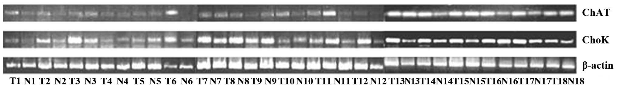
ChAT and ChoK mRNA expression in lung
cancer
RT-PCR was performed to determine the ChAT and ChoK
mRNA expression in the tumor and normal tissues. The primers used
for RT-PCR are shown in Table 1.
As shown in Fig. 1 and compared
with the expression in normal lung tissues, the mRNA expression of
ChAT increased in nine out of the 18 lung tumors, including four
adenocarcinomas, two squamous cell carcinomas, one atypical
carcinoid, one small cell carcinoma and one mucoepidermoid
carcinoma (T1, T2, T6, T8, T11, T12, T14, T16 and T17; Fig. 1). Increased mRNA expression of ChoK
was found in 14 out of the 18 lung tumors, including five
adenocarcinomas, seven squamous cell carcinomas, one atypical
carcinoid and one small cell carcinoma (T1, T2, T3, T6, T7, T8, T9,
T11, T12, T13, T14, T15, T17 and T18; Fig. 1).
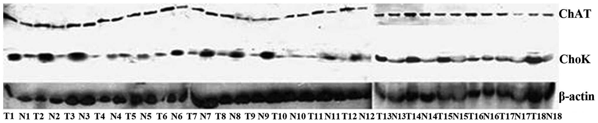
ChAT and ChoK protein expression in lung
cancer
Western blot analysis was performed to detect the
ChAT and ChoK protein expression. As compared with the expression
in the normal lung tissues, the ChAT protein expression was
upregulated in nine out of the 18 lung tumors (T1, T2, T6, T8, T11,
T12, T14, T16 and T17; Fig. 2) and
the ChoK protein expression increased in 14 out of the 18 lung
tumors (T1, T2, T3, T6, T7, T8, T9, T11, T12, T13, T14, T15, T17
and T18; Fig. 2).
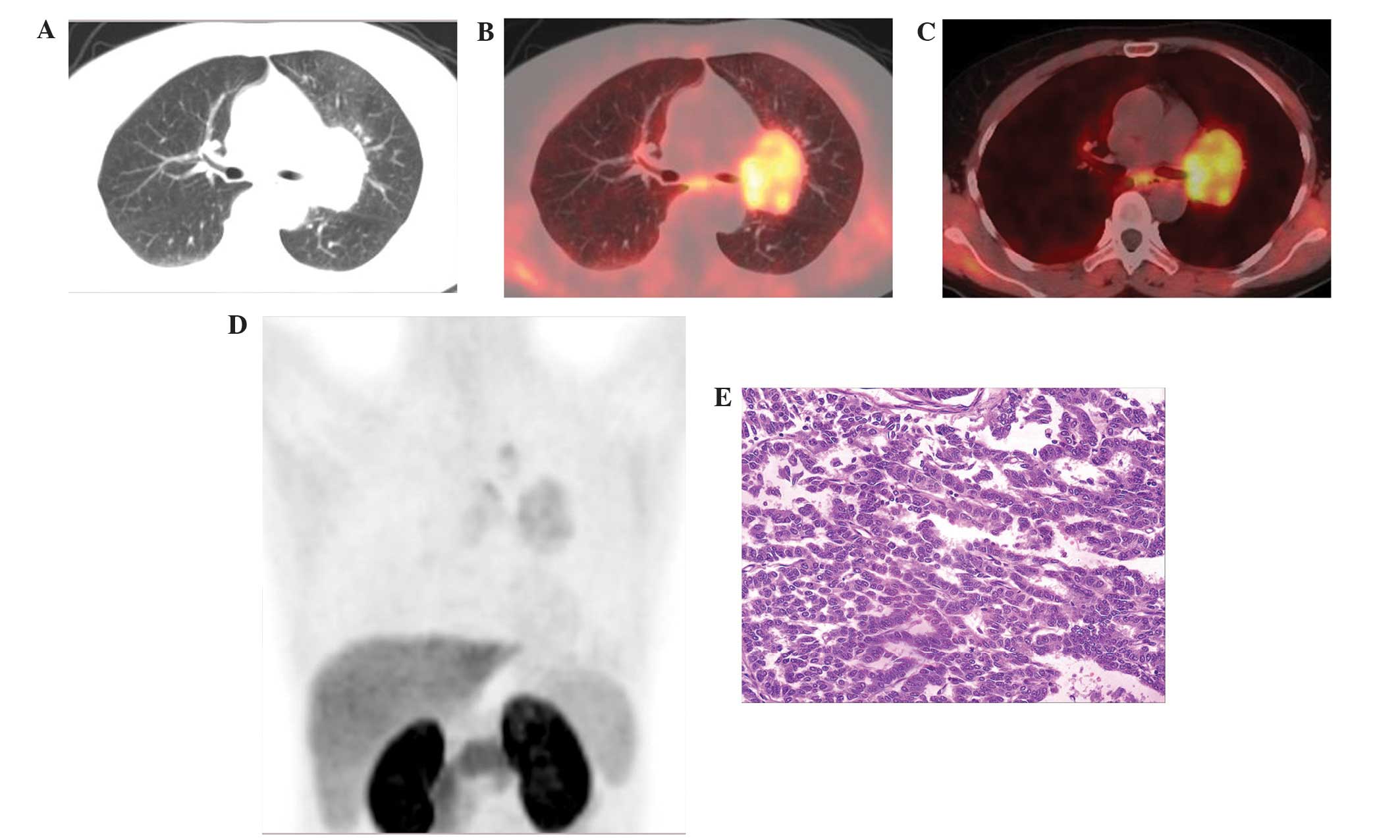
A total of eight of the 18 lung tumors exhibited a
significant increase in the mRNA and protein expression of ChoK and
ChAT, including four adenocarcinomas, two squamous cell carcinomas,
one atypical carcinoid and one small cell carcinoma. A total of
three of the 18 lung tumors (one squamous cell carcinoma, one
adenocarcinoma and one atypical carcinoid) did not exhibit
expression of ChAT and ChoK, which were assigned to group 1
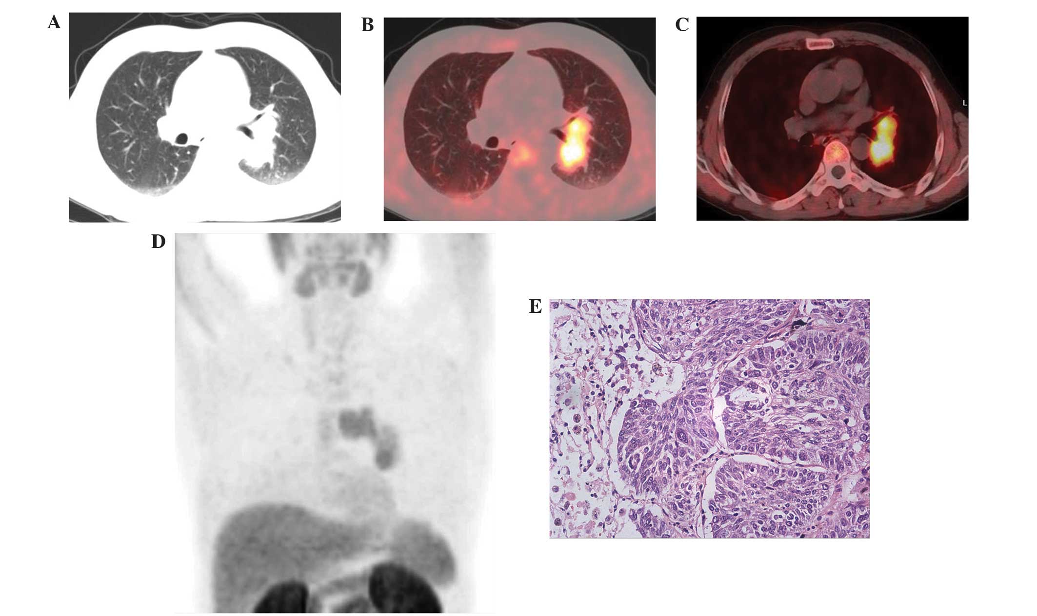
(Fig. 3). The other 15 lung tumors
with high expression of ChoK and/or ChAT were assigned to group 2
(Figs. 4 and 5).
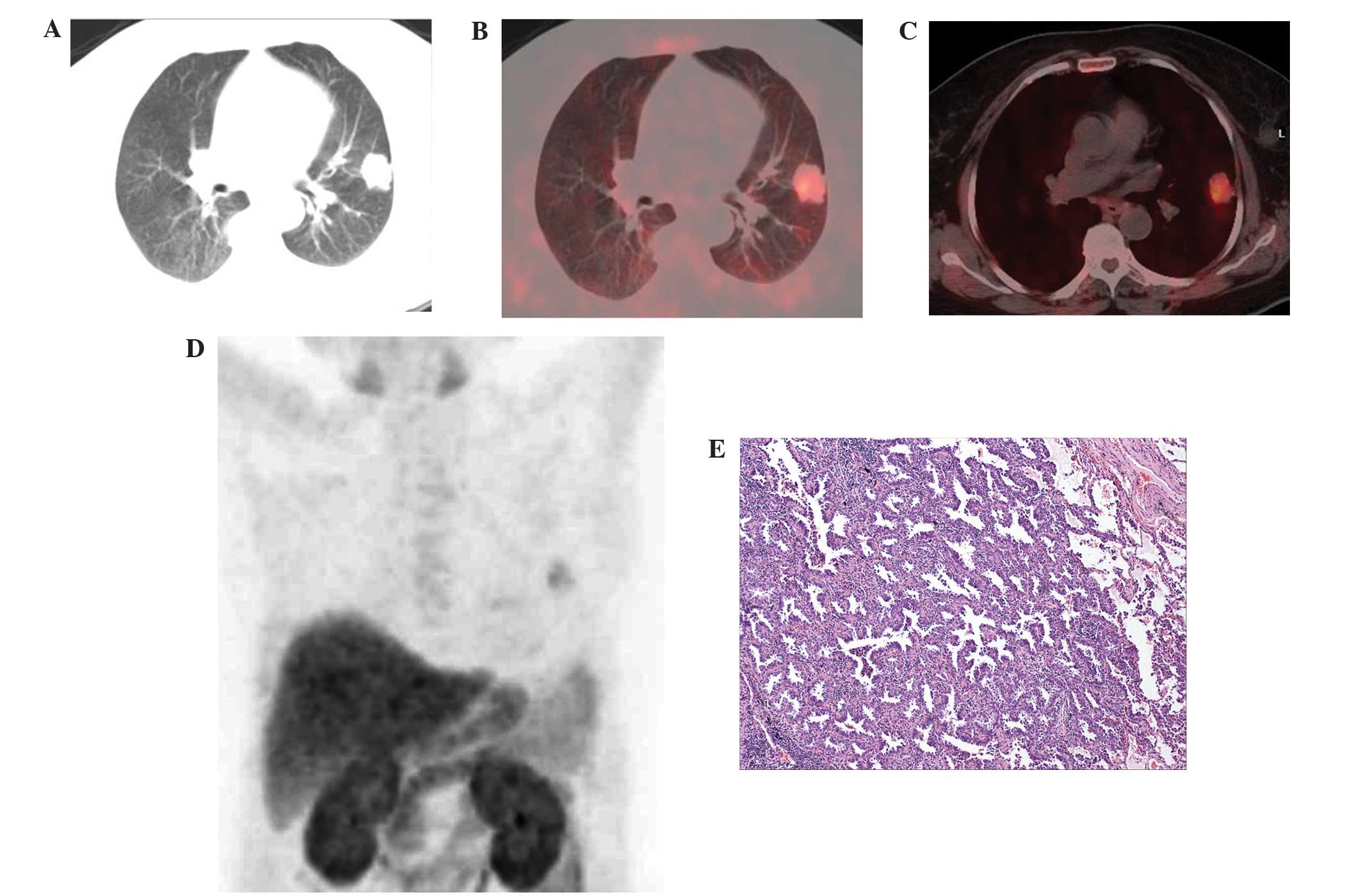
Characteristics and semiquantitative
analysis of 11C-Choline PET/CT imaging
All lung cancer lesions were visualized using the
11C-Choline PET/CT imaging (Figs. 3, 4 and 5).
PET/CT also revealed metastases in the lung hilar lymph nodes
(13/17) and mediastinal lymph nodes (10/13) with a clear focal
accumulation of 11C-Choline.
Quantification revealed that the values of SUVmean,
SUVmax and T/NT were 3.55±1.63, 4.24±1.92 and 5.78±1.85,
respectively, for all the lung tumors (data not shown). For the
eight squamous cell carcinomas, the SUVmean, SUVmax and T/NT values
were 3.61±1.90, 4.17±2.22 and 6.86±1.90, respectively and these
values for the six adenocarcinomas were 3.12±1.52, 3.80±1.94 and
4.88±1.49, respectively. The differences were not significant for
the values of SUVmean, SUVmax and T/NT between the squamous cell
carcinomas and adenocarcinomas (t=0.512, P=0.618; t=0.325, P=0.75;
t=1.987, P=0.07; respectively).
Comparative analysis did not identify a statistical
difference for the values of SUVmean, SUVmax and T/NT between
groups 1 and 2 (P=0.603, 0.577 and 0.463, respectively). The
SUVmean, SUVmax and T/NT values in group 1 were 4.01±2.14,
4.83±2.56 and 5.04±1.31, respectively and these values in group 2
were 3.46±1.58, 4.12±1.86 and 5.93±1.94, respectively (data not
shown).
Discussion
Several studies have assessed the value of
11C-Choline PET for detection and staging of lung cancer
and the results have been controversial (15–17).
The metabolism of choline in lung cancer cells has also been
investigated in several studies (10–13,18–20).
The majority of studies have suggested that phosphorylation is the
only pathway of 11C-Choline metabolism in cancer cells
(10–11), however, certain studies have
demonstrated that acetylation is another pathway of choline
metabolism in small cell lung cancer and acetylcholine acts as an
autocrine growth factor in small cell lung cancer (12,13).
In lung cancer cells, nicotinic acetylcholine receptor (nAChR)
subunit transcripts have also been observed to be expressed at
varying levels and one or more subunits are highly expressed in
non-small cell lung carcinomas (18,19).
In addition, the protein expression of nAChR subunits has been
investigated (18,20). This suggests that the acetylation
pathway is important in lung cancer.
The present study demonstrated that the mRNA and
protein expression of ChAT and ChoK increased in nine and 14 of the
18 lung tumors, respectively, compared with the expression in
normal lung tissues. A total of eight of the 18 cases exhibited a
high expression of ChAT and ChoK. The only case with high
expression of ChAT was identified to be mucoepidermoid carcinoma.
The results indicated that the phosphorylation pathway was the main
pathway of choline metabolism. There also exists an acetylation
pathway in lung cancer and the acetylation pathway may exist not
only in small cell lung cancer but also in non-small cell lung
cancer. It is reasonable to consider that the increased mRNA and
protein expression of ChAT was associated with the high expression
of nAChR in lung cancer cells.
However, three out of the 18 tumors exhibited no
expression of ChAT and ChoK, but exhibited a high uptake of
11C-Choline, which may be due to other factors,
including high expression of nAChR, the oxidation pathway or tumor
blood flow. In addition, the SUV and T/NT values between groups 1
and 2 did not demonstrate statistical significance, which was
another representation of the choline metabolism in lung cancer;
however why the case without expression of ChAT and ChoK exhibited
a high uptake of 11C-Choline remains to be elucidated.
These results require further investigation in future studies.
The present study has several limitations. Firstly,
the number of patients who enrolled in the present study was
limited. The small population may lead to biased results. Secondly,
the present study included cases of lung cancer, without benign
lesions. Certain studies have revealed that specific types of
benign lesions, including inflammatory granulation tissue exhibit a
high uptake of 11C-Choline (7,21).
Identification of the 11C-Choline metabolic mechanism in
benign lesions may assist in differentiating malignant and benign
lesions. In addition, the mRNA and protein expression of ChAT and
ChoK were investigated. Other factors, including nAChR expression,
the oxidation pathway and tumor blood flow may also have a role in
11C-Choline uptake and metabolism, which are to be
investigated in a future study.
In conclusion, all lung tumor lesions were
visualized using 11C-Choline PET/CT imaging. The
phosphorylation and acetylation pathways may be important in
11C-Choline metabolism in different pathological types
of lung cancer.
Acknowledgements
This study was supported by the National Natural
Science Foundation of China (grant nos. 81001223 and 81301868), the
Shandong Province Science and Technology Development Plan (grant
nos. 2012GSF11820, 2012YD18053 and 2012YD18086) and the Medical
Science and Technology Project of Shandong Province (grant no.
2009QW014).
References
|
1
|
American Cancer Society. Global Cancer
Facts and Figures. 2nd Edition. http://www.cancer.org/acs/groups/content/@epidemiologysurveilance/documents/document/acspc-027766.pdf.
American Cancer Society; Atlanta, GA, USA: 2011
|
|
2
|
Almuhaideb A, Papathanasiou N and Bomanji
J: 18F-FDG PET/CT imaging in oncology. Ann Saudi Med.
31:3–13. 2011. View Article : Google Scholar : PubMed/NCBI
|
|
3
|
Baum RP, Swietaszczyk C and Prasad V:
FDG-PET/CT in lung cancer: an update. Front Radiat Ther Oncol.
42:15–45. 2010. View Article : Google Scholar
|
|
4
|
Schillaci O, Travascio L, Bolacchi F,
Calabria F, Bruni C, Cicciò C, Guazzaroni M, Orlacchio A and
Simonetti G: Accuracy of early and delayed FDG PET-CT and of
contrast-enhanced CT in the evaluation of lung nodules: a
preliminary study on 30 patients. Radiol Med. 114:890–906. 2009.(In
English and Italian). View Article : Google Scholar : PubMed/NCBI
|
|
5
|
DeGrado TR, Coleman RE, Wang S, Baldwin
SW, Orr MD, Robertson CN, Polascik TJ and Price DT: Synthesis and
evaluation of 18F-labeled choline as an oncologic tracer
for positron emission tomography: initial findings in prostate
cancer. Cancer Res. 61:110–117. 2001.PubMed/NCBI
|
|
6
|
Nanni C, Rubello D and Fanti S: Could
choline PET play a role in malignancies other than prostate cancer?
Eur J Nucl Med Mol Imaging. 35:216–218. 2008. View Article : Google Scholar
|
|
7
|
Tian M, Zhang H, Oriuchi N, Higuchi T and
Endo K: Comparison of 11C-choline PET and FDG PET for
the differential diagnosis of malignant tumors. Eur J Nucl Med Mol
Imaging. 31:1064–1072. 2004.PubMed/NCBI
|
|
8
|
Picchio M, Giovannini E, Crivellaro C,
Gianolli L, di Muzio N and Messa C: Clinical evidence on PET/CT for
radiation therapy planning in prostate cancer. Radiother Oncol.
96:347–350. 2010. View Article : Google Scholar : PubMed/NCBI
|
|
9
|
Roivainen A, Forsback S, Grönroos T,
Lehikoinen P, Kähkönen M, Sutinen E and Minn H: Blood metabolism of
(methyl-11C)choline; implications for in vivo imaging
with positron emission tomography. Eur J Nucl Med. 27:25–32. 2000.
View Article : Google Scholar : PubMed/NCBI
|
|
10
|
Hara T: 11C-choline and
2-deoxy-2-(18F)fluoro-D-glucose in tumor imaging with
positron emission tomography. Mol Imaging Biol. 4:267–273. 2002.
View Article : Google Scholar
|
|
11
|
Yoshimoto M, Waki A, Obata A, Furukawa T,
Yonekura Y and Fujibayashi Y: Radiolabeled choline as a
proliferation marker: comparison with radiolabeled acetate. Nucl
Med Biol. 31:859–865. 2004. View Article : Google Scholar : PubMed/NCBI
|
|
12
|
Song P, Sekhon HS, Jia Y, Keller JA,
Blusztajn JK, Mark GP and Spindel ER: Acetylcholine is synthesized
by and acts as an autocrine growth factor for small cell lung
carcinoma. Cancer Res. 63:214–221. 2003.PubMed/NCBI
|
|
13
|
Song P, Sekhon HS, Proskocil B, Blusztajn
JK, Mark GP and Spindel ER: Synthesis of acetylcholine by lung
cancer. Life Sci. 72:2159–2168. 2003. View Article : Google Scholar : PubMed/NCBI
|
|
14
|
Pascali C, Bogni A, Itawa R, Cambie M and
Bombardieri E: [11C]Methylation on a C18
Sep-Pak cartridge: a convenient way to produce
[N-methyl-11C]choline. J Labelled Comp Rad. 43:195–203.
2000. View Article : Google Scholar
|
|
15
|
Pieterman RM, Que TH, Elsinga PH, Pruim J,
van Putten JW, Willemsen AT, Vaalburg W and Groen HJ: Comparison of
(11) C-choline and (18)F-FDG PET in primary
diagnosis and staging of patients with thoracic cancer. J Nucl Med.
43:167–172. 2002.PubMed/NCBI
|
|
16
|
Wang T, Sun YE, Yao SL, Yu CH, Yin DY and
Tian JH: Value of carbon-11 choline positron emission tomography in
patients with pulmonary abnormalities. Zhonghua Wai Ke Za Zhi.
44:405–408. 2006.(In Chinese). PubMed/NCBI
|
|
17
|
Khan N, Oriuchi N, Zhang H, Higuchi T,
Tian M, Inoue T, Sato N and Endo K: A comparative study of
11C-choline PET and (18F)fluorodeoxyglucose
PET in the evaluation of lung cancer. Nucl Med Commun. 24:359–366.
2003. View Article : Google Scholar : PubMed/NCBI
|
|
18
|
Maus AD, Pereira EF, Karachunski PI,
Horton RM, Navaneetham D, Macklin K, Cortes WS, Albuquerque EX and
Conti-Fine BM: Human and rodent bronchial epithelial cells express
functional nicotinic acetylcholine receptors. Mol Pharmacol.
54:779–788. 1998.PubMed/NCBI
|
|
19
|
Lam DC, Girard L, Ramirez R, Chau WS, Suen
WS, Sheridan S, Tin VP, Chung LP, Wong MP, Shay JW, Gazdar AF, Lam
WK and Minna JD: Expression of nicotinic acetylcholine receptor
subunit genes in non-small-cell lung cancer reveals differences
between smokers and nonsmokers. Cancer Res. 67:4638–4647. 2007.
View Article : Google Scholar : PubMed/NCBI
|
|
20
|
Zia S, Ndoye A, Nguyen VT and Grando SA:
Nicotine enhances expression of the alpha 3, alpha 4, alpha 5, and
alpha 7 nicotinic receptors modulating calcium metabolism and
regulating adhesion and motility of respiratory epithelial cells.
Res Commun Mol Pathol Pharmacol. 97:243–262. 1997.PubMed/NCBI
|
|
21
|
Zhang H, Tian M, Oriuchi N, Higuchi T,
Watanabe H, Aoki J, Tanada S and Endo K: 11C-choline PET
for the detection of bone and soft tissue tumours in comparison
with FDG PET. Nucl Med Commun. 24:273–279. 2003. View Article : Google Scholar : PubMed/NCBI
|