Introduction
Lung cancer is one of the primary causes of cancer
mortality worldwide (1). Non-small
cell lung cancer (NSCLC) accounts for ~85% of lung cancer cases, of
which 75% exhibit distant metastases at diagnosis (2,3).
Since the introduction of numerous novel chemotherapy drugs,
chemotherapy has been reported to significantly improve survival
(4) however, chemotherapeutic
toxicity has been detected in large numbers of patients (5).
Monoclonal antibodies and small molecule receptor
tyrosine kinase inhibitors (TKIs) are two targeted drugs commonly
used for treating patients with advanced NSCLC (6). Previous studies have revealed that
epidermal growth factor receptor (EGFR)-TKIs are an effective
treatment strategy that has a beneficial effect on tumors with EGFR
gene mutations (6,7). Furthermore, an increase in EGFR gene
copy numbers has been demonstrated to be associated with improved
survival prognosis for patients treated with TKI (8). In addition, 20–40% of patients with
NSCLC have concurrent mutations and gene amplification (9,10).
Recent studies have also demonstrated that some TKI-effective
patients have no significant EGFR genetic changes (11). For TKIs, there are no clear patient
selection criteria. In addition to mutation and genetic
amplification, there may be other effects of targeted drug
mechanisms (12)
MicroRNAs (miRNAs/miRs) are a class of mature small
non-coding RNAs, varying between 22 and 25 nucleotides in length,
that regulate gene expression at the post-transcriptional level by
promoting degradation of target mRNAs or inhibition of protein
synthesis (12,13). Dysregulated miRNA expression has
been demonstrated to be associated with numerous tumor types, thus
suggesting that certain miRNAs can function as oncogenes or tumor
suppressor genes (13). EGFR may
be a miRNA128-b target gene, and it has been demonstrated that in
NSCLC cells there is loss of heterozygosity in miRNA128-b, which is
associated with EGFR-TKIs curative efficacy (14). However, the effect of miRNA-128-b
on the regulation of EGFR expression in NSCLC remains unclear.
The present study aimed to investigate miRNA-128-b
and EGFR expression levels in NSCLC cancer tissue compared with
adjacent normal tissue, and investigate the association between the
two factors and clinicopathological factors in patients with NSCLC,
in order to determine the role of miRNA-128-b expression in lung
cancer and the regulation of EGFR expression.
Materials and methods
Clinical data
Between March and August 2014, tissue specimens
(cancer tissue and normal adjacent tissue) were collected following
surgical treatment of 42 patients with NSCLC in Shandong Tumor
Hospital (Jinan, China). Patients had not previously received any
other preoperative radiotherapy and chemotherapy therapies.
Following collection, tissue specimens were stored at −80°C prior
to subsequent analysis. The diagnosis and classification of these
patients were performed by a professional pathologist, and later
verified by subsequent morphological and immunohistochemistry
analyses. Patients were staged according to the TNM system
(15). The present study was
granted ethical approval by the Shandong Tumor Hospital Ethics
Committee, and written informed consent was obtained from patients.
According to the alterations in miRNA-128-b expression in cancer
tissues relative to normal tissues, tissue specimens were divided
into the following three groups: The descending group, stable group
and increasing group. The clinical data of patients included in the
present study are presented in Table
I.
 | Table I.Clinical information of patients and
microRNA-128-b expression levels in cancerous and normal
tissue. |
Table I.
Clinical information of patients and
microRNA-128-b expression levels in cancerous and normal
tissue.
|
| miRNA-128-b relative
expression |
|
|
|---|
|
|
|
|
|
|---|
| Variable | D | S | I | Percentage (%) | P-value |
|---|
| Sex |
|
|
|
| 0.89 |
| Male | 23 | 5 | 6 | 0.81 |
|
|
Female | 5 | 1 | 2 | 0.19 |
|
| Age (years) |
|
|
|
| 0.28 |
|
<60 | 16 | 3 | 2 | 0.50 |
|
| ≥60 | 12 | 3 | 6 | 0.50 |
|
| Smoking |
|
|
|
| 0.9 |
| No | 12 | 2 | 3 | 0.40 |
|
| Yes | 16 | 4 | 5 | 0.60 |
|
| Drinking |
|
|
|
| 0.17 |
| No | 20 | 2 | 6 | 0.67 |
|
| Yes | 8 | 4 | 2 | 0.33 |
|
| TNM stage |
|
|
|
| 0.72 |
| I | 9 | 2 | 1 | 0.29 |
|
| II | 5 | 2 | 2 | 0.21 |
|
| III | 14 | 2 | 5 | 0.50 |
|
| Pathological
type |
|
|
|
| 0.9 |
|
Adenocarcinoma | 10 | 3 | 3 | 0.38 |
|
|
Squamous | 11 | 2 | 4 | 0.40 |
|
|
Others | 7 | 1 | 1 | 0.21 |
|
| Histology
grade |
|
|
|
| 0.38 |
|
Low | 3 | 1 | 3 | 0.17 |
|
|
Medium | 20 | 4 | 5 | 0.69 |
|
|
High | 5 | 1 | 0 | 0.14 |
|
| EGFR mRNA |
|
|
|
| <0.01 |
| D | 3 | 2 | 8 | 0.31 |
|
| I | 25 | 4 | 0 | 0.69 |
|
| EGFR protein
expression |
|
|
|
| 0.03 |
|
Positive | 8 | 5 | 5 | 0.60 |
|
|
Negative | 11 | 0 | 1 | 0.40 |
|
Cell culture
Lung adenocarcinoma cell line A549 was obtained from
the American Type Culture Collection (Manassas, VA, USA), cultured
in RPMI containing 10% fetal bovine serum (Hyclone; GE Healthcare,
Chicago, IL, USA) at 37°C, and subsequently maintained in a 5%
CO2 humidification incubator.
miRNA transfection
Cells (1.0–1.5×10⁵) were seeded into
6-well plates and after 24 h miRNA molecules (miRNA-128-b,
5′-UCACAGUGAACCGGUCUCUUU-3′; miRNA-128-b inhibitor,
5′-AAAGAGACCGGUUCACUGUGA-3′; mimics NC,
5′-UUGUACUACACAAAAGUACUG-3′; inhibitor NC,
5′-CAGUACUUUUGUGUAGUACAA-3′; Guangzhou RiboBio, Co., Ltd.,
Guangzhou, China) were transfected into cells using Lipofectamine
2000 (Invitrogen; Thermo Fisher Scientific, Inc.), and the final
plasmid concentration was 150 nM. Cells were collected 24 h
post-transfection for subsequent RNA extraction and drug
sensitivity analysis, and 48 h post-transfection for immunostaining
analyses.
RNA extraction
Total RNA extraction was performed using TRIzol
reagent (Invitrogen; Thermo Fisher Scientific, Inc., Waltham, MA,
USA), and isolated RNA samples were then treated with DNase (2
U/µg; Promega Corporation, Madison, WI, USA).
Reverse transcription
(RT)-quantitative polymerase chain reaction (qPCR) analysis of
miRNA-128-b
A mirVana Quantitative RT-PCR miRNA kit (Ambion;
Thermo Fisher Scientific, Inc.) was used to perform RT (44°C for 1
h and 92°C for 10 min) according to the manufacturer's protocol.
Briefly, total RNA (200 ng) was added to specific RT primers to
synthesize complementary (c)DNA. The qPCR reaction consisted of the
initial denaturation at 95°C for 10 min, followed by 40 cycles for
15 sec at 95°C and 1 min at 60°C. The expression of miR128-b
relative to RNA, U6 small nuclear 1 (RNU6B) was calculated using
the 2−ΔΔCq method (16). All assays were performed in
triplicate. Primer sequences were as follows: miRNA-128-b, forward,
5′-CGCGCTCACAGTGAACCG-3, reverse, 5′-GTGCAGGGTCCGAGGT-3′; and
RNU6B, forward, 5′-CTCGCTTCGGCAGCACA-3′ and reverse,
5′-AACGCTTCACGAATTTGCGT-3′.
Semi-quantitative RT-PCR
RT was performed using ProtoScript® cDNA
single-stranded synthesis kit (New England Biolabs, Inc., Ipswich,
MA, USA), according to the manufacturer's protocol; PCR
amplification was performed using Platinum Blue PCR SuperMix
reagent (Invitrogen; Thermo Fisher Scientific, Inc.). Initial
denaturation was performed at 94°C for 2 min followed by 40 cycles
of 94°C for 20 sec and 56°C for 55 sec; and a final extension step
for 1 min at 72°C. mRNA primer sequences used were as follows: EGFR
forward, 5′-ATGCTCCCACCAC-3′ and reverse,
5′-CCCTTCGCACTTCTTACAC-3′; and β-actin forward,
5′-ATGATGATATCGCCGCGCT-3′ and reverse, 5′-TGGGTCATCTTCTCGCGGTT-3′.
The amplified product was analyzed on a 1.25% agarose gel, and EGFR
mRNA expression was determined by grayscale scanning of the agarose
gel containing ethidium bromide. Expression levels were calculated
relative to β-actin expression. Data were semi-quantified using
LightCycler analysis software v1.50 (Roche Diagnostics, Mannheim,
Germany). All experiments were performed in triplicate.
EGFR immunohistochemistry (IHC)
analysis
IHC was performed to investigate the expression of
EGFR protein. After blocking the non-specific protein-binding sites
with 20% goat serum (OriGene Technologies, Inc., Beijing, China)
for 20 min at room temperature, anti-EGFR antibody staining
(1:1,000; cat. no. PA1-1110; Zymed; Thermo Fisher Scientific, Inc.)
was performed on A549 cells that were fixed with 4%
paraformaldehyde at room temperature for 15 min and tissue sections
obtained from patients that were fixed with 4% paraformaldehyde at
4°C (17). Subsequently, they were
incubated with a goat anti-rabbit immunoglobulin-horseradish
peroxidase secondary antibody (1:200; cat. no. TA130023; OriGene
Technologies, Inc.) at 37°C for 30 min. Tissue sections were
classified as follows: 0, <10% positive cells; 1, 10% positive
cells; 2, 11–50% positive cells; 3, >50% positive cells. The
number of positive cells per field of view (0–100%) and staining
intensity (0, negative; 1 weak positive; 2, medium positive; 3,
strong positive) of EGFR was determined (18). Images were captured using an
IX71-SIF microscope (Olympus Corporation, Tokyo, Japan) at ×200
magnification. Two independent observers blinded to tissue status
determined staining intensities in cell cytoplasm. Image-Pro Plus
software (Media Cybernetics, Inc., Rockville, MD, USA) was used to
quantify the mean densities of the expression intensities of EGFR,
as previously described (18).
Immunohistochemical positive staining in the cytoplasm was
assessed, and two observers blinded to tissue status evaluated
positive staining of each tissue core. An average of five images
per tissue core was used for quantitative analysis.
Statistical analysis
All experiments were repeated three times and the
data are presented as the mean ± standard deviation. Statistical
significant differences were determined by one-way analysis of
variance followed by Fisher's least significant difference post-hoc
test. Qualitative data were correlated using the Pearson
correlation. Rates were compared using the χ2 test.
Statistical analysis was performed using SPSS 19.0 software (IBM
Corp., Armonk, NY, USA). P<0.05 was considered to indicate a
statistically significant difference.
Results
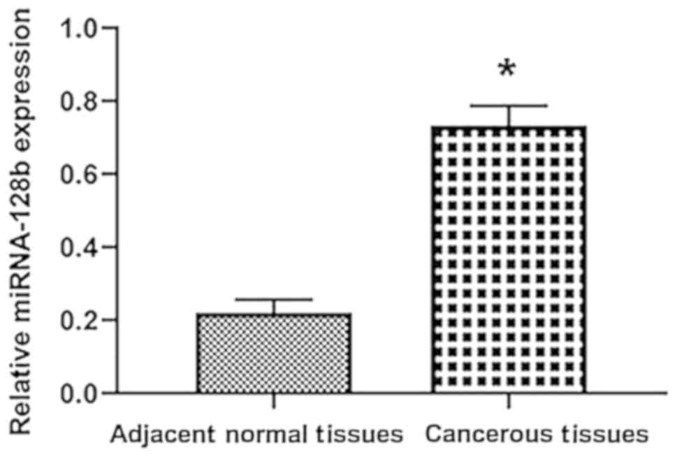
miRNA-128-b expression in tissues
RT-qPCR was performed to detect the expression of
miRNA-128-b in the cancerous tissues and adjacent normal tissues of
42 patients, and the results revealed that expression was not
detected in 16 cancerous tissues or in 8 adjacent normal tissues.
In 6 patients, miR-128-b expression was not detected in either
cancerous or adjacent tissues (Fig.
1). The expression of miRNA 128-b in 28 cases (66.7%) of 42
cases was suppressed compared with normal tissues [tumor tissue
(T): Normal tissue (N) <1]. The mean value of miRNA 128-b
expression in cancer tissues was 0.2±0.08, and the mean value of
miRNA 128-b expression in normal tissues was 0.7±0.16 (P=0.04).
Association between miRNA-128-b
expression and clinical pathological factors
Relative expression levels of miRNA128-b was
revealed to be significantly associated with differences in EGFR
mRNA expression and EGFR protein expression. However, no
significant differences in miRNA 128-b expression regarding
differences in sex, age, pathological type, operation stage and
degree of differentiation were determined (Table I).
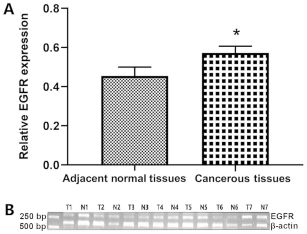
EGFR mRNA expression in tissues
RT-PCR was performed to detect EGFR mRNA expression
in the cancer tissues and normal tissues of 42 patients (Fig. 2). The results revealed that EGFR
mRNA expression was enhanced in cancer tissue compared with normal
tissue in 69% of patients (T:N≥1.0). The mean EGFR mRNA expression
was revealed to be 0.57±0.16 in the cancer tissue, and 0.48±0.16 in
normal tissue (P=0.02).
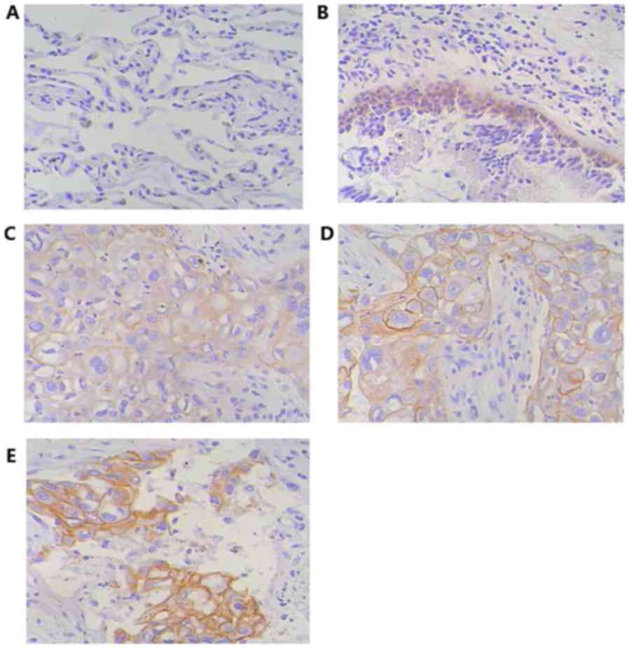
EGFR protein expression in tissue
IHC staining of EGFR protein was performed in 30 of
the 42 tissue samples. EGFR protein staining was revealed to be
negative (Fig. 3A) or weakly
positive (Fig. 3B) in normal
tissue samples, in which staining was limited to basal levels
(<10% positive cells). EGFR protein expression in cancer tissues
was revealed to be increased compared with normal tissue samples.
When the number of positive cells was 10% (1), the sample was considered positive for
EGFR protein expression; EGFR positive expression was detected in
18 (60%) cancer tissues (Fig. 3C).
When the number of positive cells was >10% (2 and 3), the sample
was considered to have high EGFR protein expression; EGFR protein
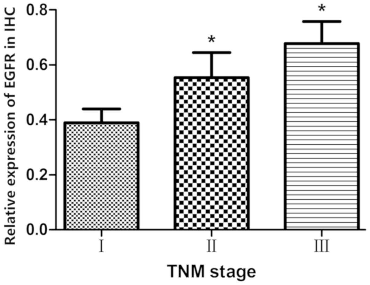
was highly expressed in 10 patients (Fig. 3D and E). As demonstrated in
Fig. 4, EGFR protein expression
was associated with TNM stage (P<0.05 TNM stage II or III vs.
TNM stage I).
Association between the expression of
miRNA-128-b and the expression of EGFR in lung cancer tissues
Pearson correlation analysis demonstrated that there
were significant negative correlations between the relative
expression of miRNA128-b and both EGFR mRNA relative expression and
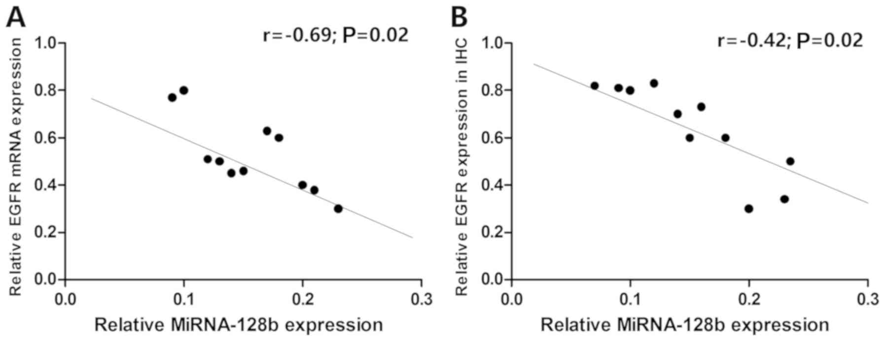
EGFR protein expression (r=−0.69, P=0.02, Fig. 5A; r=−0.43, P=0.02, Fig. 5B).
miRNA-128-b regulates EGFR expression
in lung cancer cell lines
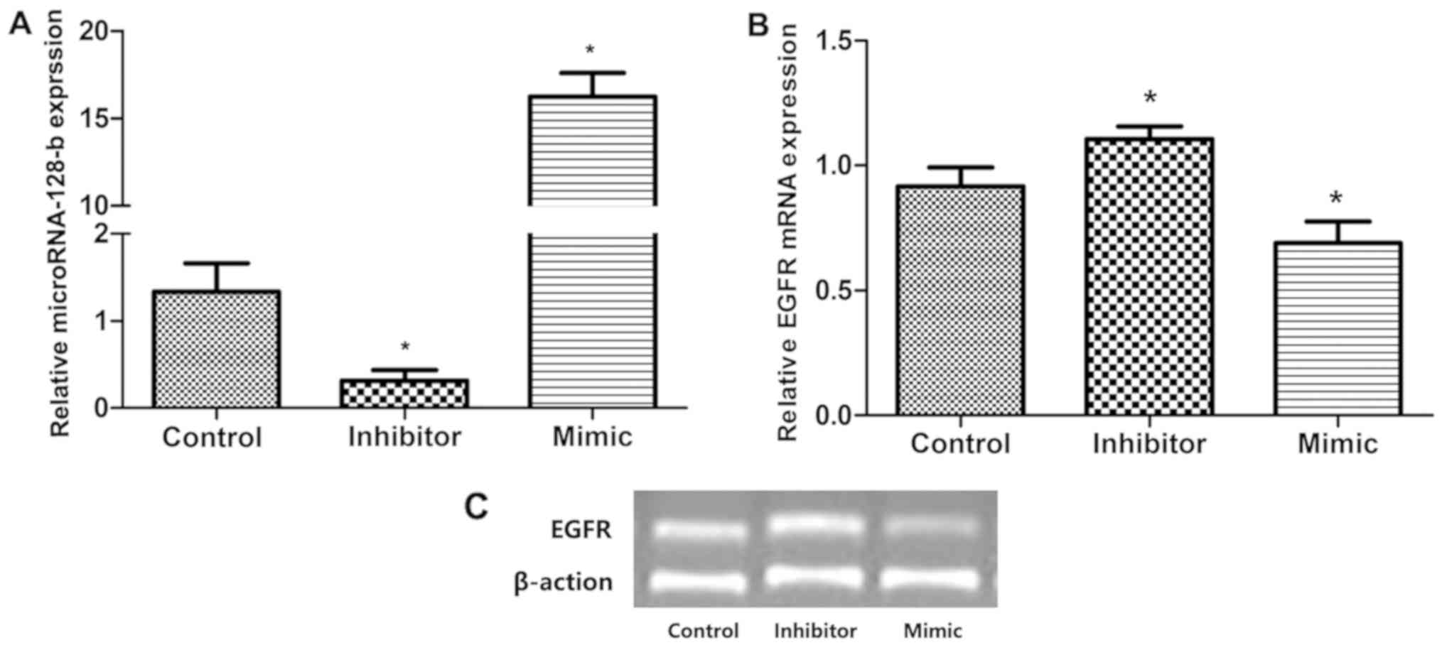
miRNA-128-b mimics and inhibitors were transfected
into A549 cells expressing EGFR protein to investigate the effect
of miRNA-128-b on the regulation of EGFR expression. The expression
of miRNA-128-b in cells transfected with mimics significantly
increased compared with the control group, and the inhibitor group
exhibited significantly decreased miRNA-128-b expression levels
compared with the control group. Compared with the control group,
EGFR mRNA expression was significantly suppressed in cells
transfected with miRNA-128-b mimics compared with control cells;
whereas EGFR mRNA expression was significantly increased the
inhibitor-treated cells compared with control cells (P<0.05;
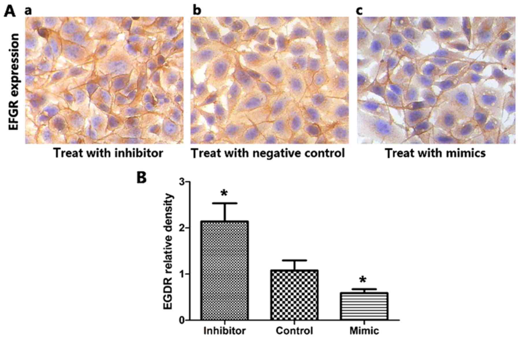
Fig. 6A and B). In addition,
Fig. 7 presented the results of
immunohistochemistry analysis following transfection with
miRNA-128-b mimics or inhibitors. The expression of EGFR protein in
cells treated with miRNA-128-b inhibitors was significantly
upregulated compared with control cells; whereas EGFR protein
expression was significantly downregulated in cells treated with
miRNA-128-b mimics compared with control cells (P<0.05 vs.
control; Fig. 7B).
Discussion
Over the past 40 years, the understanding of
potential cellular or genetic changes associated with lung cancer
has developed. The EGFR signal transduction pathway has been
suggested to represent a therapeutic target, leading to the
development of small-molecule TKIs. It has been reported that
EGFR-TKIs are only effective in 10–30% of patients with
chemotherapy-refractory NSCLC (19–21),
and so the identification of novel regulatory molecules may
optimize targeting therapy for lung cancer.
miRNAs are a class of small molecule non-coding
RNAs, and a considerable number of microRNAs are located in
tumor-associated gene loci or fragile sites, thus suggesting that
miRNAs may be involved in tumorigenesis. The deletion of chromosome
3p allele is common in the early stages of lung cancer, which
results in dysregulation of EGFR expression (18). miRNA-128-b is located on chromosome
3p22, and regulates EGFR expression in NSCLC cells via binding with
the 3′ untranslated region (3′UTR) (18).
To the best of our knowledge, the association
between miRNA-128-b expression and EGFR expression has not been
reported in NSCLC, the previous study conducted a systematic
analysis (10). EGFR may be a
miRNA-128-b target gene, and it has been demonstrated that in NSCLC
cells there is loss of heterozygosity in miRNA-128-b, which is
associated with EGFR-TKIs curative efficacy (20,21).
Firstly, differences in miRNA-128-b expression and EGFR expression
between cancerous tissue and normal tissue were compared.
miRNA-128-b expression was occasionally not detected in tumor
tissues and was revealed to be decreased in cancerous tissue
compared with normal tissue. The results demonstrated that EGFR
mRNA was expressed in all samples; however, the expression of EGFR
mRNA was revealed to be enhanced in cancerous tissue compared with
normal tissue. The difference of the same mRNA expression between
normal tissues and cancerous tissues may be a result of the
heterogeneity of the expression patterns in different tissue types
or due to the biological behavior of the tumor. IHC results
demonstrated that the expression of EGFR in normal tissues was
negative or weakly positive, whereas the expression of EGFR protein
in cancerous tissues was increased compared with normal tissues.
Consistent with a previous study (22), expression levels of EGFR protein
were revealed to be increased in stage III NSCLC compared with
earlier stages of NSCLC. It has been previously suggested that EGFR
expression increases progressively in a stage-dependent manner in
NSCLC (23). In addition, the
correlation between miRNA128-b expression and EGFR expression was
analyzed in cancerous tissues. Pearson correlation analysis
demonstrated that there were significant negative correlations
between the relative miRNA128-b expression and relative EGFR mRNA
and protein expression
Previous studies have investigated the association
between miRNA expression and mRNA expression in NSCLC tissues.
Tumor suppressor candidate 2 (TUSC2) is a tumor suppressor gene and
is located on chromosome 3p21. miRNA-93 and miRNA-197 bind to the
TUSC2 3′UTR to negatively regulate its expression in NSCLC tissues
(24). The results of the present
study demonstrated that the relative expression of miRNA-128-b was
negatively correlated with the relative expression of EGFR mRNA in
cancerous tissue. In addition, the results revealed that
miRNA-128-b expression was sometimes absent in the tumor compared
with the normal tissue, and the relative expression of miRNA-128-b
was decreased, unchanged and increased in three cases in cancer
tissues, and was significantly correlated with EGFR protein
expression.
Furthermore, the results of the present study
suggested that miRNA-128-b regulates EGFR expression in NSCLC
cells. A549 cells expressing EGFR and miRNA-128-b were
investigated, and EGFR expression was revealed to be downregulated
following transfection with mimics, whereas EGFR expression was
upregulated following treatment with inhibitors, and drug
sensitivity was subsequently increased. Similarly, H157 cells are
EGFR-expressing cell lines, and a previous study revealed that
treatment with miRNA-128-b inhibitors upregulated EGFR protein
expression, while EGFR protein expression was downregulated
following miRNA-128-b mimic treatment (18).
The results of present study suggested that
miRNA-128-b expression and EGFR expression were differentially
expressed in cancer tissues compared with normal tissues, and the
relative expression of miRNA-128-b was negatively correlated with
EGFR mRNA and EGFR protein expression levels in cancerous tissues.
miRNA-128-b regulated EGFR expression in NSCLC cells, thereby
affecting the drug sensitivity of cells. These results suggested
that miRNA-128-b may exhibit an inhibitory role in lung cancer.
However, the efficacy of existing anti-EGFR drugs for the treatment
of lung cancer is limited, and thus the development of novel
therapeutic strategies is necessary to successfully suppress signal
transduction and EGFR expression. Inhibition of signal transduction
via EGFR-TKIs and inhibition of EGFR expression via miRNA-128-b may
represent a novel therapeutic strategy for the treatment of
patients with EGFR mutant-harboring NSCLC.
Acknowledgements
Not applicable.
Funding
The Natural Science Foundation of Shandong Province
(grant no. Y2005C49) supported the present study.
Availability of data and materials
All data generated or analyzed during this study are
included in this published article.
Authors' contributions
LL and DW conceived and designed the study. DW
collected the data. LL performed the experiments, prepared the
diagrams and drafted the manuscript. LL and DW reviewed and edited
the manuscript. All authors read and approved the final
manuscript.
Ethics approval and consent to
participate
The present study was granted ethical approval by
the Shandong Tumor Hospital Ethics Committee. Written informed
consent was obtained from the patients.
Patient consent for publication
Written informed consent was obtained from the
patients.
Competing interests
The authors declare that they have no competing
interests.
References
|
1
|
Kligerman S and White C: Epidemiology of
lung cancer in women: Risk factors, survival, and screening. AJR Am
J Roentgenol. 196:287–295. 2011. View Article : Google Scholar : PubMed/NCBI
|
|
2
|
Yang JC, Kang JH, Mok T, Ahn MJ,
Srimuninnimit V, Lin CC, Kim DW, Tsai CM, Barraclough H, Altug S,
et al: First-line pemetrexed plus cisplatin followed by gefitinib
maintenance therapy versus gefitinib monotherapy in East Asian
patients with locally advanced or metastatic non-squamous non-small
cell lung cancer: A randomised, phase 3 trial. Eur J Cancer.
50:2219–2230. 2014. View Article : Google Scholar : PubMed/NCBI
|
|
3
|
Brown T, Boland A, Bagust A, Oyee J,
Hockenhull J, Dundar Y, Dickson R, Ramani VS and Proudlove C:
Gefitinib for the first-line treatment of locally advanced or
metastatic non-small cell lung cancer. Health Technol Assess. 14
(Suppl):71–99. 2010. View Article : Google Scholar : PubMed/NCBI
|
|
4
|
Mendoza L: Targeted therapies in the
treatment of advanced non-small-cell lung cancer: Update. Klin
Onkol. 22:131–138. 2009.PubMed/NCBI
|
|
5
|
Avery EJ, Kessinger A and Ganti AK:
Therapeutic options for elderly patients with advanced non-small
cell lung cancer. Cancer Treat Rev. 35:340–344. 2009. View Article : Google Scholar : PubMed/NCBI
|
|
6
|
Ahn HK, Choi YL, Han JH, Ahn YC, Kim K,
Kim J, Shim YM, Um SW, Kim H, Kwon OJ, et al: Epidermal growth
factor receptor mutation and treatment outcome of mediastinoscopic
N2 positive non-small cell lung cancer patients treated with
neoadjuvant chemoradiotherapy followed by surgery. Lung Cancer.
79:300–306. 2013. View Article : Google Scholar : PubMed/NCBI
|
|
7
|
Tanaka K, Hida T, Oya Y, Oguri T, Yoshida
T, Shimizu J, Horio Y, Hata A, Kaji R, Fujita S, et al: EGFR
mutation impact on definitive concurrent chemoradiation therapy for
inoperable stage III adenocarcinoma. J Thorac Oncol. 10:1720–1725.
2015. View Article : Google Scholar : PubMed/NCBI
|
|
8
|
Dahabreh IJ, Linardou H, Kosmidis P,
Bafaloukos D and Murray S: EGFR gene copy number as a predictive
biomarker for patients receiving tyrosine kinase inhibitor
treatment: A systematic review and meta-analysis in non-small-cell
lung cancer. Ann Oncol. 22:545–552. 2011. View Article : Google Scholar : PubMed/NCBI
|
|
9
|
Fukuoka M, Wu YL, Thongprasert S,
Sunpaweravong P, Leong SS, Sriuranpong V, Chao TY, Nakagawa K, Chu
DT, Saijo N, et al: Biomarker analyses and final overall survival
results from a phase III, randomized, open-label, first-line study
of gefitinib versus carboplatin/paclitaxel in clinically selected
patients with advanced non-small-cell lung cancer in Asia (IPASS).
J Clin Oncol. 29:2866–2874. 2011. View Article : Google Scholar : PubMed/NCBI
|
|
10
|
Murphy M and Stordal B: Erlotinib or
gefitinib for the treatment of relapsed platinum pretreated
non-small cell lung cancer and ovarian cancer: A systematic review.
Drug Resist Updat. 14:177–190. 2011. View Article : Google Scholar : PubMed/NCBI
|
|
11
|
Pirker R: What is the best strategy for
targeting EGF receptors in non-small-cell lung cancer. Future
Oncol. 11:153–167. 2015. View Article : Google Scholar : PubMed/NCBI
|
|
12
|
Cheng Q, Yi B, Wang A and Jiang X:
Exploring and exploiting the fundamental role of microRNAs in tumor
pathogenesis. Oncol Targets Ther. 6:1675–1684. 2013.
|
|
13
|
Xia H and Hui KM: MicroRNAs involved in
regulating epithelial-mesenchymal transition and cancer stem cells
as molecular targets for cancer therapeutics. Cancer Gene Ther.
19:723–730. 2012. View Article : Google Scholar : PubMed/NCBI
|
|
14
|
Weiss GJ, Bemis LT, Nakajima E, Sugita M,
Birks DK, Robinson WA, Varella-Garcia M, Bunn PA Jr, Haney J,
Helfrich BA, et al: EGFR regulation by microRNA in lung cancer:
Correlation with clinical response and survival to gefitinib and
EGFR expression in cell lines. Ann Oncol. 19:1053–1059. 2008.
View Article : Google Scholar : PubMed/NCBI
|
|
15
|
Chheang S and Brown K: Lung cancer
staging: Clinical and radiologic perspectives. 30:99–113.
2013.PubMed/NCBI
|
|
16
|
Yanaihara N, Caplen N, Bowman E, Seike M,
Kumamoto K, Yi M, Stephens RM, Okamoto A, Yokota J, Tanaka T, et
al: Unique microRNA molecular profiles in lung cancer diagnosis and
prognosis. Cancer Cell. 9:189–198. 2006. View Article : Google Scholar : PubMed/NCBI
|
|
17
|
Wang W, Wen Q, Xu L, Xie G, Li J, Luo J,
Chu S, Shi L, Huang D, Li J and Fan S: Activation of Akt/mTOR
pathway is associated with poor prognosis of nasopharyngeal
carcinoma. PLoS One. 9:e1060982014. View Article : Google Scholar : PubMed/NCBI
|
|
18
|
Ling LI and Song LH: The role of MicroRNA
in lung cancer. J Clin Oncol. 36:1377–1380. 2009.(In Chinese).
|
|
19
|
Zhao N, Zhang XC, Yan HH, Yang JJ and Wu
YL: Efficacy of epidermal growth factor receptor inhibitors versus
chemotherapy as second-line treatment in advanced non-small-cell
lung cancer with wild-type EGFR: A meta-analysis of randomized
controlled clinical trials. Lung Cancer. 85:66–73. 2014. View Article : Google Scholar : PubMed/NCBI
|
|
20
|
Wang S, Su X, Bai H, Zhao J, Duan J, An T,
Zhuo M, Wang Z, Wu M, Li Z, et al: Identification of plasma
microRNA profiles for primary resistance to EGFR-TKIs in advanced
non-small cell lung cancer (NSCLC) patients with EGFR activating
mutation. J Hematol Oncol. 8:1272015. View Article : Google Scholar : PubMed/NCBI
|
|
21
|
Duan X and Shi J: Advance in microRNAs and
EGFR-TKIs secondary resistance research in non-small cell lung
cancer. Zhongguo Fei Ai Za Zhi. 17:860–864. 2014.(In Chinese).
PubMed/NCBI
|
|
22
|
Aichler M, Motschmann M, Jütting U, Luber
B, Becker K, Ott K, Lordick F, Langer R, Feith M, Siewert JR and
Walch A: Epidermal growth factor receptor (EGFR) is an independent
adverse prognostic factor in esophageal adenocarcinoma patients
treated with cisplatin-based neoadjuvant chemotherapy. Oncotarget.
5:6620–6632. 2014. View Article : Google Scholar : PubMed/NCBI
|
|
23
|
Ludovini V, Bellezza G, Pistola L,
Bianconi F, Di Carlo L, Sidoni A, Semeraro A, Del Sordo R,
Tofanetti FR, Mameli MG, et al: High coexpression of both
insulin-like growth factor receptor-1 (IGFR-1) and epidermal growth
factor receptor (EGFR) is associated with shorter disease-free
survival in resected non-small-cell lung cancer patients. Ann
Oncol. 20:842–849. 2009. View Article : Google Scholar : PubMed/NCBI
|
|
24
|
Du L, Schageman JJ, Subauste MC, Saber B,
Hammond SM, Prudkin L, Wistuba II, Ji L, Roth JA, Minna JD and
Pertsemlidis A: miR-93, miR-98, and miR-197 regulate expression of
tumor suppressor gene FUS1. Mol Cancer Res. 7:1234–1243. 2009.
View Article : Google Scholar : PubMed/NCBI
|