Introduction
Menstruation is a phenomenon unique to females in
which vaginal bleeding occurs due to endometrial shedding; this
phenomenon commonly occurs in humans, most primates, and other
animals (1,2). A normal menstrual cycle in humans
consists of proliferative, secretory, and menstrual phases with two
peaks of oestrogen secretion and one peak of progesterone
secretion. Investigation of the menstrual cycle is a rapidly
emerging area of research in reproductive physiology; however, the
limited availability of menstruating animals, including higher
order primates, elephant shrews, and bats, for scientific research
remains a challenge (3–5). Furthermore, the use of these animals
to study menstrual physiology is limited owing to their low
fertility rates, high feeding cost, and ethical consideration
(6). A recent study showed that
the spiny mouse is the only naturally menstruating rodent. However,
this North African native animal is vulnerable to environmental
influences that affect normal menstruation, and its use for
menstrual studies is limited due to its unique physiology, and the
fact that it lacks antigens and antibodies for its immune response
(7). Therefore, commonly used
laboratory animals such as mice, which have a high rate of
reproduction and clear genetic background, are suitable for in
vivo research. Furthermore, they are inexpensive to procure and
well-suited for the development of a menstrual model. The present
study has reviewed the development of a mouse model of menstruation
and its research application, and described the mechanisms of
menstruation. A mouse model may be used to simulate various
gynaecological complications caused by menstrual disorders in
women, including menstrual pain and abnormal uterine bleeding.
Platelet-activating factor (PAF) is known to aggravate menstrual
pain, and activin serves a role in endometrial repair. These
findings complement and validate the results of studies on
gynaecological symptoms caused by menstrual abnormalities in
females. Therefore, a mouse model of menstruation may provide a
strong theoretical foundation for studies on the treatment of
reproductive and gynaecological diseases in humans.
Mouse model of menstruation
For a long time, previous studies had not attempted
to develop a mouse model of menstruation, because mice do not
menstruate under normal conditions. However, studies began to
investigate new animal models for experimental research to improve
understanding of gynaecological diseases. In 1940, Christiaens
(8) first transplanted human
endometrium into the anterior chamber of a macaque monkey.
Subsequently, studies began to investigate the possibility of using
the most commonly available mouse to study menstruation (9). Consequently, rodent animals were
introduced as experimental models to investigate menstruation.
In the 1960s, a mouse model was used to study the
mechanism of menstruation (10).
In 1984, two distinguished reproductive scientists, C. A. Finn and
M. Pope, excised ovaries from mice and treated them with hormones;
after they induced decidualization by using oil, they
subcutaneously injected progesterone; and by removing progesterone
from the mouse endometrium, a mouse endometrial breakdown model was
constructed for the first time (11). In 2003, Brasted et al
(12) applied a progesterone
implant to the mouse menstruation model generated by Finn and Pope.
The initiation of progesterone withdrawal was optimized following
decidual induction and the model was improved. In 2007, Xu et
al (13) used mifepristone (a
progesterone receptor antagonist) to block progesterone and induced
uterine decidualisation to successfully develop a mouse model of
menstruation. In 2009, Kaitu'u-Lino et al (14) used wild-type (WT) mice and mice
overexpressing follistatin (a natural activin inhibitor) to study
menstruation. In this study, the uterus was excised and cultured
in vitro following progesterone withdrawal; subsequently,
the human endometrial epithelial ECC-1 cell line was used to
simulate repair. This study provided a novel theoretical basis for
the clinical treatment of abnormal uterine bleeding. In 2010,
Kikuchi-Arai et al (15)
used severe combined immunodeficiency (SCID)/γCnull (NOG) mice to
study menstruation. Following the excision of ovaries, human
endometrial fragments were injected subcutaneously into the back of
the mice, and periodic hormone therapy was applied to develop an
immunodeficient mouse model of the menstrual cycle. This study
demonstrated that uterine natural killer (NK) cells do not
originate from the peripheral blood, but from the endometrium. In
2014, Cousins et al (16)
used a non-surgical embryo transfer device (NSET) to inject sesame
oil into the uterine cavity of mice to induce endometrial
decidualization, and intraperitoneally injected bromodeoxyuridine
prior to sacrifice to develop a modified mouse model of menstrual
repair. In the same year, an endometriosis mouse model based on the
menstrual model was developed (17). To construct this model, ovaries
were excised from mice and periodic hormone therapy was used to
induce menstruation. During menstruation, menstrual endometrial
tissue of a model with the same genetic background were inoculated
into the peritoneum of immunocompetent mice, causing endometriosis.
In 2017, De Clercq et al (18) used conventional methods to develop
mouse models of menstruation. In this study, transient receptor
potential (TRP) channel expression in uterine horns was measured
using reverse transcription-quantitative polymerase chain reaction
at different time points following discontinuation of progesterone
administration to identify factors promoting mouse embryo
implantation. In 2018, Peterse et al (19) demonstrated that laparoscopic
injection-induced uterine decidualisation is greater than that
induced by vaginal injection. Ovariectomized mice were treated with
cyclic hormones, following which oil was injected into the uterus
using laparoscopy, laparotomy, and vaginal methods. This study
demonstrated an optimised method to induce uterine decidualisation
in a mouse model of menstruation. In the same year, Hellman et
al (20) used WT and
platelet-activating factor (PAF)-knockout mice to induce
menstruation using conventional methods. In this study, carbamyl
PAF (CPAF) and prostaglandin F2α (PGF2α) were intraperitoneally
injected during menstruation to compare the level of menstrual pain
in mice. In 2019, Wang et al (21) demonstrated that the placement of a
menstruating mouse model of artificial decidualisation into
restraint tubes decreases luteinizing hormone, follicle stimulating
hormone, and progesterone levels in mice under stress, and results
in endometrium breakdown and shedding. This study highlighted the
effect of pressure on menstrual regulation. Other mouse models of
menstruation continue to be investigated.
Methods of constructing a hormone induction
model
Specific induction of a menstruation model is a
complex processs. To begin with, the three requirements for
menstruation, hormonal preparation cycle, endometrial
decidualisation and progesterone withdrawal, need to be understood
(13,22). Multiple models have been developed
and improved over the years. In a study by Finn and Pope in 1984
(9), mice ovariectomized under
anaesthesia were allowed to recover for one week and subsequently
subjected to the following hormone therapy: Intraperitoneal
injection of 100 ng oestradiol on days 1 and 2; no hormone
administration on days 3, 4, and 5; and injection of 500 µg
progesterone and 10 ng oestradiol (each dissolved in 50 µl arachis
oil) on days 6 and 7. Simultaneously, 20 µl arachis oil was
injected into the uterine horn of 84 mice treated with the
aforementioned hormones. Of these, 70 (83%) exhibited
decidualisation and marked iris reaction was observed in their
uterine horns on day 2 post-injection. In addition, stromal
changes, including dilation and congestion of blood vessels,
swelling of red blood cells, rupture of blood vessel walls, and
blood exudation were observed. These endometrial changes were
similar to those observed during menstruation, indicating
successful induction of menstruation in mice (11).
Xu et al improved Brasted's model-building
method (12,13). Mice were allowed to recover for two
weeks after ovariectomy, and were injected with 100 ng 17-B
oestradiol (aromatic oil) subcutaneously on days 1, 2, and 3. Mice
were administered 50 ng progesterone and 5 ng 17B-E2 in aromatic
oil on day 7. On day 9, 20 µl arachis oil was injected into the
left corner of the mouse uterus to induce decidualisation. The mice
were sacrificed at 0, 8, 16, 24, 32, 40, and 48 h following
mifepristone administration. Unlike Finn and Pope, Xu et al
optimized the initiation of progesterone withdrawal following the
induction of decidualisation and effectively used mifepristone for
the first time.
These are two typical methods for constructing mouse
menstrual models, which are presented in Fig. 1. Details on the construction of
other mouse menstrual models are listed in Table I. The menstrual cycle in mice is
similar to that in humans. To begin with, the mice were
ovariectomized, the interference of ovarian hormones on the model
construction was eliminated, and estrogen was injected (23). The endometrium of the mice began to
thicken, and the glands and blood vessels proliferated. After a few
days, with the simultaneous injection of progesterone and estrogen,
the endometrium and glands continued to grow under hormonal action
and secreted mucus to prepare for fertilized egg implantation.
Injection of peanut oil into the mouse uterine cavity stimulated
the decidualization of the endometrium. Finally, after the
progesterone implant was removed, with the rapid decline of
hormones, endometrial blood vessels began to spasm; the endometrium
became necrotic due to ischemia; and blood and endometrial debris
flowed out of the mouse's vagina, forming menstrual blood (24,25).
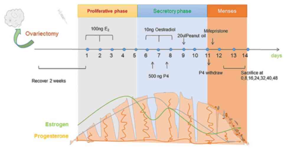 | Figure 1.Development of a mouse model of
menstruation. The mice underwent ovariectomy and were allowed to
recover for 2 weeks. Thereafter, they were administered 100 ng
oestradiol (stored in peanut oil) on days 1, 2, and 3. The mice did
not receive any treatment on days 4 and 5. Subsequently, 10 ng
oestradiol and 500 ng progesterone were injected into the uterus on
days 6, 7, and 8, and oestradiol was injected on day 9. Next, 20 µl
peanut oil is injected to induce uterine decidualisation. After two
days, mifepristone was administered to induce progesterone
withdrawal and menstruation in mice. The mice were sacrificed at 0,
8, 16, 32, 40, and 48 h after progesterone withdrawal, and the
uterine horn was obtained. |
 | Table I.Comparison of the similarities and
differences between different mouse menstrual models. |
Table I.
Comparison of the similarities and
differences between different mouse menstrual models.
| Type stage | Year | Ovary removal | Hormone
treatment | Induced
decidualization |
Post-processing | Significance | (Refs) |
|---|
| Preliminary | 1984 | Yes | Cycle | Oil injected into
the | Mice were
sacrificed and | Developed
mouse | (11) |
| exploration |
|
| therapy | uterus | uteri were
harvested | endometrial |
|
| of basic |
|
|
|
|
| rupture model |
|
| menstrual | 2003 | Yes | Cycle | Sesame oil
injected | Mice were
sacrificed at the | Optimized | (12) |
| models |
|
| therapy | into the
uterine | time of implant
removal 0 | progestin |
|
|
|
|
|
| horn | and 12, 16, 20, 24,
36, and 48 h | withdrawal
time |
|
|
| 2007 | Yes | Cycle | Arachis oil
was | Mice were
sacrificed | Mifepristone | (13) |
|
|
|
| therapy | injected into
the | at different time
points | was first used
as |
|
|
|
|
|
| left uterine
horn | following
administration of | a progesterone |
|
|
|
|
|
|
| mifepristone | withdrawal |
|
|
| 2014 | Yes | Cycle | Sesame seed
oil | Mice received an
intra- | A modified
mouse | (16) |
|
|
|
| therapy | was inserted
into | peritoneal
injection of BrdU | menstrual
repair |
|
|
|
|
|
| the uterine by | 90 min before
being | model |
|
|
|
|
|
| NSET | sacrificed |
|
|
|
| 2018 | Yes | Cycle therapy | Oil was injected in
the uterus via laparotomy, laparoscopy or vagina | Progesterone
withdrawal, followed by hysterectomy after 4 to 6 h | Identified the best
way to induce uterine decidualization | (19) |
| Improvement | 2009 | Yes | Cycle | Sesame oil was | Mice were
sacrificed 24 | Discover
possible | (14) |
| of menstrual |
|
| therapy | injected into
the | or 48 h after
progesterone | targets for
treating |
|
| model to |
|
|
| lumen of the
right | removal and the
uterus was | abnormal
uterine |
|
| simulate |
|
|
| uterine horn | removed for in
vitro analysis | bleeding |
|
| clinical |
|
|
|
| and simulation of
repair |
|
|
| disease | 2014 | Yes | Cycle | Oil was
injected | The endometrium
of | Successful | (17) |
|
|
|
| therapy | into the
uterine | artificially
induced | induction of |
|
|
|
|
|
| horn | menstruation was
transferred | endometriosis |
|
|
|
|
|
|
| to the peritoneum
of recipient | based on a |
|
|
|
|
|
|
| mice to induce
endometriosis | menstrual
model |
|
|
| 2017 | Yes | Cycle | Vaginal intra- | Uteri were
harvested at | Study the | (18) |
|
|
|
| therapy | uterine injection
of | different time
points to | factors
affecting |
|
|
|
|
|
| sesame oil | study the
expression of TRP | mouse embryo |
|
|
|
|
|
|
| channels | implantation |
|
|
| 2018 | Yes | Cycle | Oil was
injected | Intraperitoneal
injection | Improvement of | (20) |
|
|
|
| therapy | into the
uterine | of CPAF and
PGF2 | menstrual pain |
|
|
|
|
|
| horn | during artificially
induced | research based
on |
|
|
|
|
|
|
| menstruation in
mice | a menstrual
model |
|
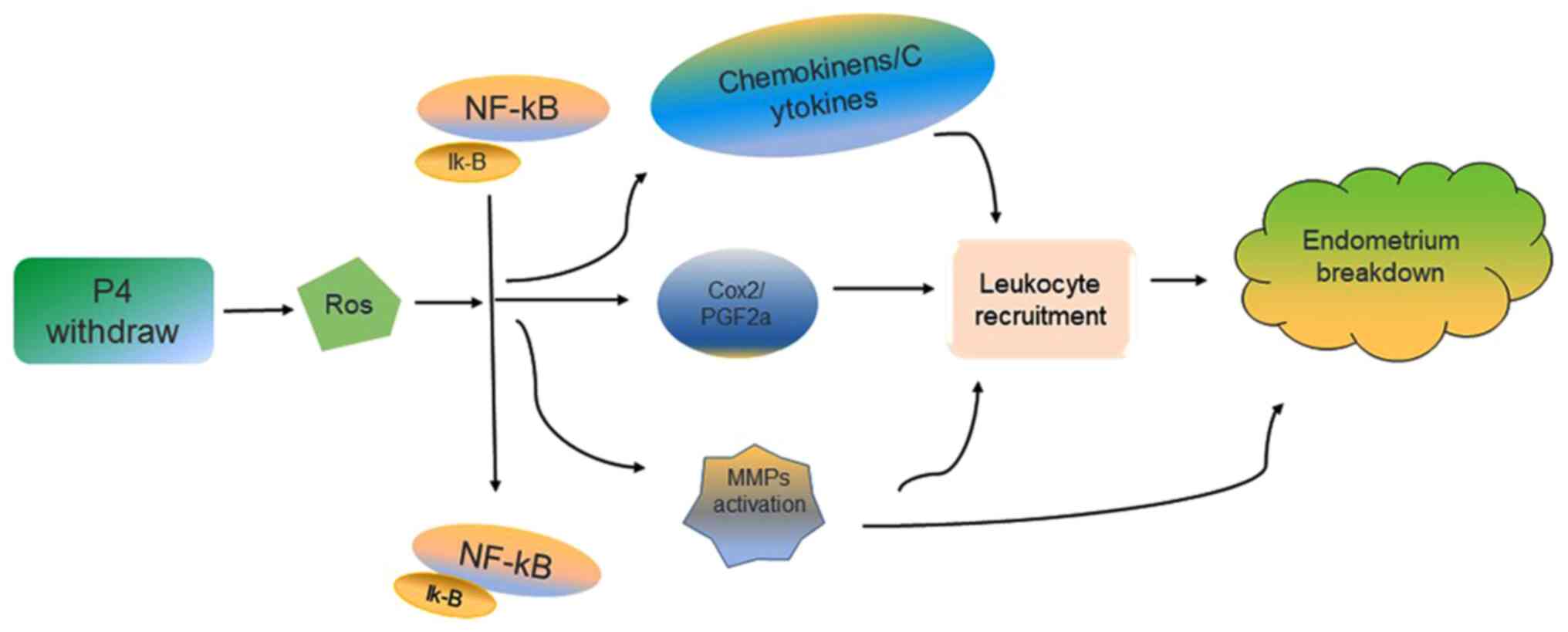
Role of NF-κB signalling pathways in
endometrial shedding
Progesterone withdrawal may lead to endometrial
breakdown and loss during the establishment of a mouse model.
However, the physiological significance of downstream processes,
reactive oxygen species (ROS), and other antioxidants in the female
reproductive system are not known. ROS have been reported to serve
an important role in menstruation (Fig. 2). Wu et al have developed a
mouse model of menstruation based on the method of Brasted et
al (12,13). In this model, the reactive oxygen
scavenger, N-acetylcysteine (NAC), was intraperitoneally
injected at different doses prior to discontinuation of
progesterone administration. The ROS assay and histological
analysis following uterine collection demonstrated that ROS levels
may affect endometrial breakdown. NAC, p65, and p50 may affect
NF-κB activation by regulating its nuclear and cytoplasmic
expression. In addition, several leukocyte chemokines are regulated
by NF-κB, the source of the beginning of ROS (26). The Cox-2 promoter binds to NF-κB
and regulates gene transcription. The in vitro, in vivo, and
molecular analysis results of NF-κB-Cox-2 signal transduction, as
well as the antioxidant NAC, has been used to determine the role of
ROS in menstruation; ROS are the key regulators of endometrial
breakdown as they regulate the downstream NF-κB signal (27).
MMPs may be broken down during menstruation, and
MMP9 is the first component that is degraded (28). In addition to ROS, RU486 may induce
endometrial breakdown. After the progesterone hormone level was
reduced, NF-κB was activated, which induced the expression of the
downstream target genes (includings MMP9) and binding to the
promoter of MMP9, further promoting the breakdown and shedding of
the mouse endometrium. The mouse model of menstruation may also be
used to determine the kinetics of endometrial breakdown and
regeneration during endometrial reconstruction (26).
Initiation of menstruation
Endometrial breakdown
The endometrium is the layer that forms the inner
lining of the uterus in mammals. It is markedly affected by the
cyclic changes in oestrogen and progesterone levels, and undergoes
proliferation, differentiation, breakdown, and repair
simultaneously (29). The
construction of the mouse model mimics this process. A rapid
decrease in progesterone levels may lead to a series of molecular
changes, including spiral artery constriction; increased production
of inflammatory cells, including white blood cells, neutrophils,
and prostaglandins; and MMP activation, leading to breakdown and
disintegration of the endometrium (15,30).
Progesterone withdrawal is an important event that initiates
endometrial breakdown and shedding, and promotes cell-factor and
factor-factor interactions. The mouse model of menstruation is a
reliable tool to understand the molecular mechanism of endometrial
breakdown.
In order to morphologically confirm the induction of
menstruation, mice are subjected to vaginal smear testing and eosin
staining following discontinuation of progesterone administration.
The presence of erythrocytes and degeneration of decidual stroma in
the vaginal smear are suggestive of menstruation. While nuclear
rupture or constriction may be clearly observed, cytoplasmic
degeneration and cytoplasmic boundary resolution are not obvious.
The uterine horn shows considerable hypertrophy and hyperemia
compared with that in the control group, in which the uterine horn
is pale pink. Blood clots, similar to those noted during human
menstruation, may be observed in the uterus. However, no spiral
artery remodeling is observed during menstruation in induced mice,
which is slightly different from human menstruation (Table II) (6,24,31).
| Table II.Comparison of human menstruation and
mouse model menstruation. |
Table II.
Comparison of human menstruation and
mouse model menstruation.
|
Characteristics | Human | Mouse model |
|---|
| Reproductive
tract | Simplex uterus with
fundal body and fallopian tubes | Bicornuate uterus
with uterine horn oviducts |
| Menstrual
cycle | Natural (28
days) | Artificially
induced |
| Breeding
season | Continuous | Continuous |
| Length of
menses | 5–7 days | 5–7 days |
| Shedding | Menstrual
waves | Menstrual
waves |
|
Decidualisation | Sponstaneous | Artificially
induced |
| Decidua
characteristics | Gentleness and
regularity | Rapid and
destructive |
| Glands | Epithelial and
intrauterine | Epithelial and
intrauterine |
| Oestrogen peak | During
proliferative and secretory phase | Artifician
induction |
| Spiral artery
remodelling | Yes | No |
| Immune
response | Yes | Yes |
Regarding menstruation, it has been proposed that P4
causes two stages of after withdrawal: P4-dependent and
P4-independent (32). The critical
period of progesterone withdrawal is earlier than the breakdown and
shedding of the endometrium in the menstrual model. However,
whether or not there is an association between endometrial
breakdown and decidual status remains unknown. The decidual state
of the mouse model is different from that in humans. In the mouse
model, reticular fibre staining and histomorphological analysis
were used to evaluate endometrial status. During endometrial
shedding, the predecidual-like zone (PZ) is actively degraded
(33). Furthermore, inhibition of
hypoxia-inducible factor-1 alpha (HIF-1α) confirmed its role in
endometrial breakdown; HIF-1α may regulate vascular endothelial
growth factor (VEGF) expression (Fig.
3). Therefore, regulation of VEGF mRNA levels may be considered
an early factor to modulate endometrial breakdown. However, HIF-1
may regulate the expression of VEGF mRNA for a long time (34). Hypoxia and HIF-1α activation are
induced by progesterone withdrawal, and hypoxia may upregulate MMP
expression. However, hypoxia is not essential for endometrial
breakdown (35).
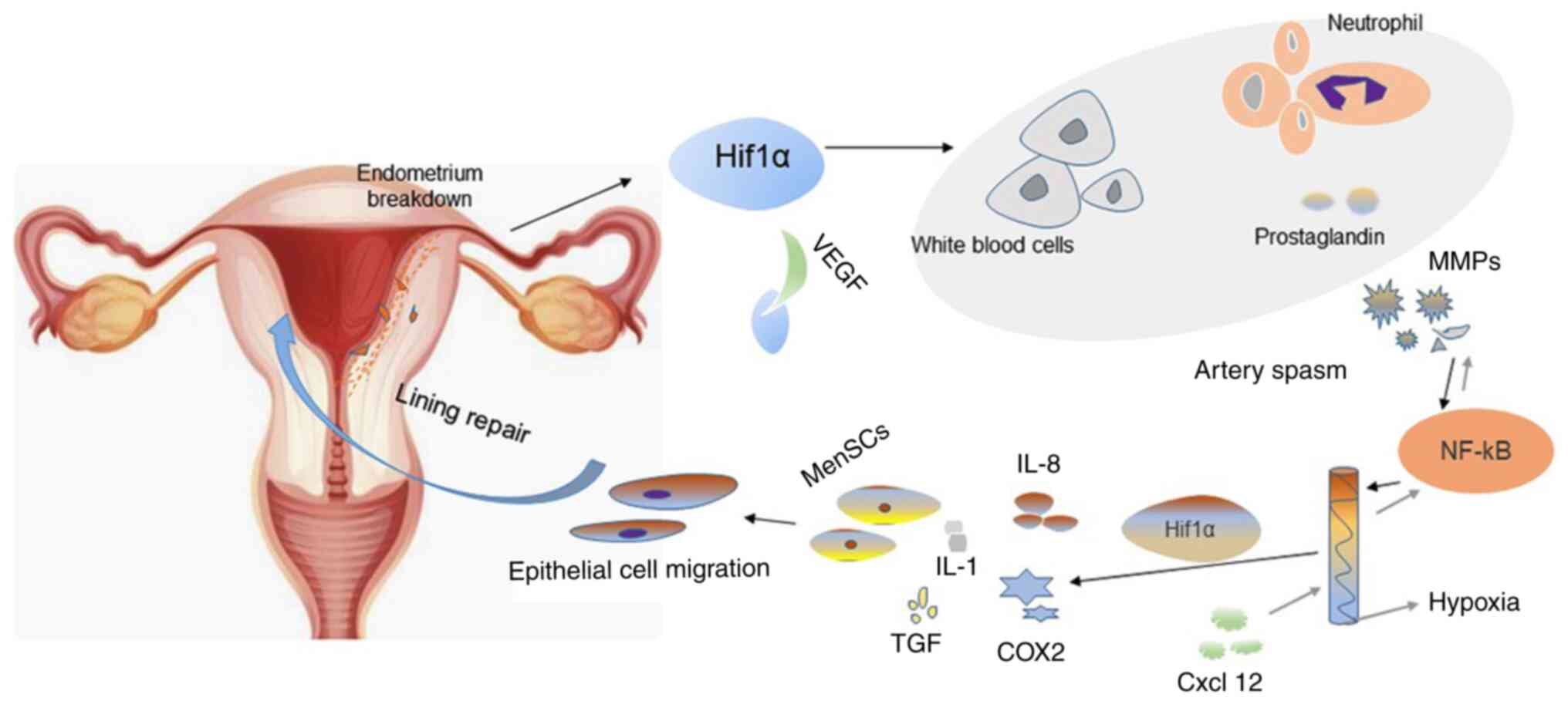 | Figure 3.Cellular and molecular changes during
endometrial breakdown and repair. Progesterone withdrawal may
promote endometrial shedding and cause menstruation. The binding of
activated HIF-1α to VEGF angiogenic factors in the endometrial
during endometrial breakdown results in increased production of
white blood cells, neutrophils, inflammatory cells, and
prostaglandins. The activation of NF-kB causes the decline of MMPs,
and the process is reversible. Spiral arteriolar muscular
contraction leads to anoxia in the endometrium during endometrial
breakdown. Endometrial repair under anaerobic conditions may result
in increased expression of IL-8 and Cox2 as well as increased
leukocyte infiltration of cytokines (e.g., IL-1) and growth factors
(e.g., transforming growth factor). The increase in the number of
menstrual blood-derived cells and migration of epithelial cells
further promotes rapid repair of the endometrium. HIF-1α,
hypoxia-inducible factor-1 α; VEGF, vascular endothelial growth
factor; MMPs, matrix metalloproteinases; IL, interleukin; Cox-2,
cyclooxygenase 2. |
Repair regulation
In humans, each menstrual cycle exhibits a unique
pattern of tissue damage, followed by rapid tissue repair without
scar tissue formation (36). For
understanding repair regulation, at present, two different methods
of tissue shedding are mainly used to induce mouse models. One of
the most commonly used models is the first proposed by Finn and
PoPe, with was later modified by Salamonsen et al to
optimize the time of endometrial shedding repair (12,23,37).
In the 1970s, a study demonstrated that epithelial
cells from the exposed end of glands and stromal tissue surface may
proliferate simultaneously, suggesting their involvement in
endometrial repair (38). The
mouse models used to study the complete endometrium repair
mechanism showed that the dynamics of the rapid healing of
endometrial serve a supporting role in the cycle,
epithelial-mesenchymal transition and epithelial cell migration
were observed 4–12 h after progesterone withdrawal (16). In a recent study by Patterson et
al (39), female mice were
mated with vasectomized male mice to induce pseudopregnancy.
Thereafter, the female mice were oophorectomized and subjected to
progesterone withdrawal to induce menstruation. The number of cells
co-expressing keratin and vimentin were increased by two-fold 24 h
after ovariectomy (39). These
results indicated that epithelial-mesenchymal transition and
epithelial cell migration may greatly enhance endometrial repair,
providing a new basis for the treatment of abnormal endometrial
repair.
Constriction of the uterus and decidual spiral
arterioles during menstruation forms a hypoxic uterine environment.
Furthermore, it is not known whether oxygen deprivation may affect
endometrial repair. Under normoxia, cell sensors may hydroxylate
certain proline residues in HIF-1α (40). In the mouse menstrual model,
bleeding and physiological anoxia of the endometrium occur. HIF-1α
has also been found to alleviate hypoxia-induced delayed
endometrial repair in mice via pharmacological activity.
Furthermore, hyperoxia may delay endometrial repair by
downregulating HIF-1α (41).
Cousins et al (42)
recorded uterine breakdown and repair over a 24-h period in mice
and measured oxygen tension in the uterus in real time. This study
reported that the number of epithelial cells varied under hypoxia.
In addition, spatiotemporal variations and changes in the
expression of VEGF and the angiogenesis gene encoding
matrix-derived factor, a receptor for chemokine ligand (CXCL12)
mRNA regulation, may affect hypoxia. These results suggested that
hypoxia-induced gene regulation during endometrial rupture is
similar to that during menstruation (42). Nonetheless, hypoxia and ischemia
caused by spiral artery spasm following progesterone withdrawal may
cause endometrial breakdown. However, hypoxia did not induce
endometrial shedding and repair 21 days after endometrial
transplantation in ovariectomized severe combined immunodeficient
mice. This suggested that hypoxia may not serve a role in
endometrial breakdown and repair in xenograft mouse models
(43).
A recent study demonstrated that the absence of
neutrophils may significantly inhibit uterine reconstruction of
menarche in a mouse model of menstruation (44). Furthermore, inflammatory mediators
and granulocytes may promote local tissue remodelling of the uterus
(45). White blood cells are
required in tissue repair 4 to 5 days prior to menstruation in
females; furthermore, progesterone withdrawal may upregulate
inflammatory mediators, including NF-κB, monocyte chemoattractant
peptide 1 (MCP-1), interleukin 8 (IL-8), and cyclooxygenase 2
(Cox-2) (46).
Previous studies have reported that MMPs (specific
proteases that bind to all components of the extracellular matrix)
serve an important role in menstruation (47,48).
In a mouse model of endometrial repair, the uterus was removed at
different time points following progesterone withdrawal and MMP
expression was measured. The results demonstrated that MMP
expression in the endometrium was significantly increaseed during
the menstrual and pre-menstrual periods than during other stages of
the menstrual cycle. However, MMP inhibitors (batimistat and
doxycycline) had no significant effect on endometrial repair in
mice; therefore, MMPs may not be the key mediators of endometrial
breakdown and repair (49).
Androgens serve an important role in endometrial repair in
menstruating mice. The administration of a single dose of androgen
may cause spatiotemporal changes in the expression of caspase-3 and
MMP3 or MMP9. Furthermore, androgen receptor (AR)-dependent
regulation of MMPs is known to accelerate endometrial tissue
repair. Therefore, AR may be a potential drug target for abnormal
endometrial repair (50).
Transcriptional regulation of these factors during endometrial
repair is important to understand the mechanism of endometrial
repair.
Applications of a mouse model of
menstruation
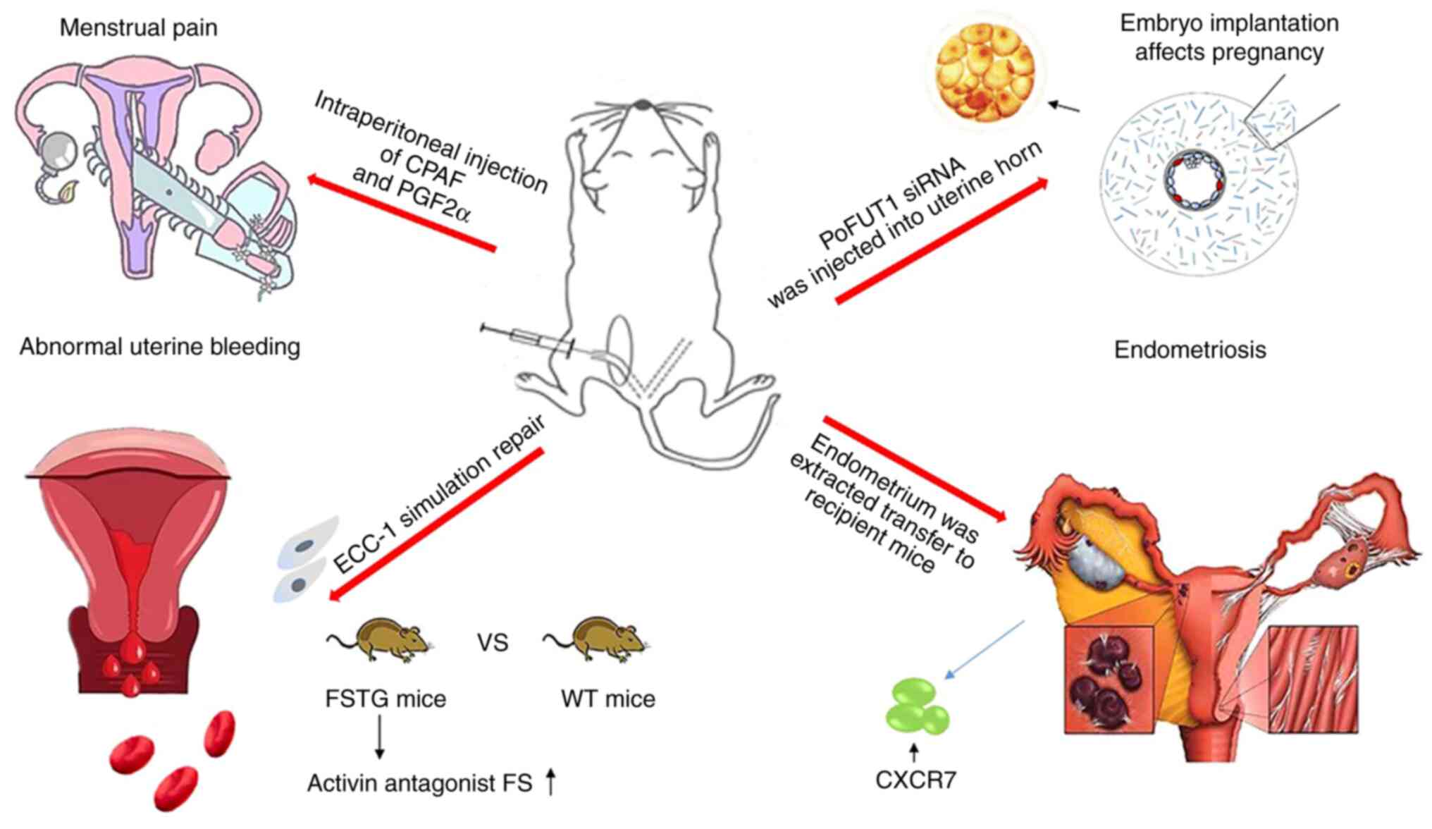
Menstrual pain
Although the development and precise mechanism of
normal menstruation are vaguely understood, pathological changes
occurring during abnormal menstruation are not yet known. The
development of a mouse model of menstruation may provide valuable
insight into various gynaecological symptoms or diseases. Endocrine
disorders may lead to menstrual disorders, including dysmenorrhoea
and abnormal bleeding (51,52).
Dysmenorrhoea is one of the most common gynaecological conditions.
Approximately 40–70% of females of reproductive age, both students
and working professionals, are significantly affected by the
adverse effects of menstrual pain. Furthermore, menstrual pain may
cause other pelvic diseases (53).
The mechanism and aetiology of menstrual pain is
different. Mice with menstrual pain have increased levels of
prostaglandin F2α (PGF2α; Table
III) (54).
Platelet-activating factor (PAF) levels are not significantly
affected by non-steroidal anti-inflammatory drugs. PAF receptor
agonist CPAF and PGF2α may increase intrauterine pressure. WT and
mice treated with the PAF receptor knockout agent were used to
construct a menstrual model, and several parameters, including
stretching, rubbing abdomen on the floor, nongrooming licking, and
arched posture were implemented to assess the pain level of mice.
Intraperitoneal injection of CPAF and PGF2α into WT mice during
menstruation caused pelvic hyperalgesia and visceral pain.
(Fig. 4) However, no pain was
detected in PAF receptor-knockout mice. CPAF may cause ischemia by
causing endometrial contraction, which may affect menstrual pain in
mice. In addition, CPAF may reduce oxygen saturation and aggravate
hypoxia in the uterus (20).
Furthermore, injection of sildenafil into the uterus of rats in the
oestrous phase increased blood flow to the uterus and decreased
dysmenorrhoea. However, the effects of ischemia and hypoxia on
uterine physiology remain controversial. Steady-state electrode
recording often ignores the effect of contraction (55). The use of mouse models may provide
a new experimental basis for developing novel targets to study
uterine contractile force, inflammatory precursors, and tissue
oxygenation.
| Table III.Changes and roles of cytokines at the
pathological level. |
Table III.
Changes and roles of cytokines at the
pathological level.
| Clinical
category | Molecular
pathology | Impact | Change |
|---|
| Menstrual pain | PGF2a | Acceleration of
endometrial contraction | Increased |
|
| CPAF | Aggravation of
anoxia | Increased |
| Abnormal uterine
bleeding | Follistatin | Promotion of
repair | Decreased |
|
|
DuP-697/indomethacin | Suppression of
menstruation | Decreased |
|
| Cox-2 | Inflammatory
response | Increased |
| Endometriosis | CXCR7 | Induction of
endometriosis | Increased |
|
| K:F9 | Promotion of
activity | Increased |
|
| KLF13 | Participation in
the pathological process | Decreased |
|
| TFF3 | Differential
marker | Increased |
|
| Iron deposit |
Erythrocytosis/excess iron storage | Increased |
| Successful embro
implantation | ESC decidua | Secretion of
factors | Increased |
|
| poFUT1 | Promotion of
endometrial decentralization | Increased |
|
| TRP | Embryo
implantation | Increased |
|
| Tiam1 | Regulation of
decidua | Increased |
|
| Omega-3 | Facilitation of
embryo implantation | Increased |
|
| MSX1 | Control of
embryonic development | Increased |
Abnormal uterine bleeding
Abnormal uterine bleeding is caused by insufficient
or incorrect repair of the endometrium following menstruation, and
it is often accompanied by initial cell damage and white blood cell
cascade inflammation (14,47).
The role of activin in promoting wound healing in
mouse endometrium has been confirmed previously (56). Therefore, WT mice and mice
overexpressing follistatin (a natural inhibitor of activin) were
used to develop a hormone therapy-induced endometrial
breakdown/repair model in ovariectomized mice. Furthermore, ECC-1
cells were used to simulate endometrial repair, and activin was
shown to promote wound closure. Furthermore, the inhibitory effect
of follistatin on endometrial repair in mice overexpressing
follistatin was significantly slower than that in WT mice,
suggesting that activin may promote endometrial repair (14).
In 2016, Cousins et al (50) constructed a mouse menstrual model.
Following progesterone withdrawal, androgens were injected, and
androgens had a significant effect on the regulation of persistent
vaginal bleeding in mice (50).
Furthermore, beta-alpha and beta-beta immunolocalization were
performed on the endometrium of mice, and staining of certain white
blood cells and specific epithelium was detected adjacent to the
repair site of the endometrium. The localization of the endometrial
repair site in the mouse model was the same as that in humans
during menstruation, with an influx of numerous white blood cells
(57). These findings may aid in
understanding the pathological mechanism of abnormal uterine
bleeding.
Cyclooxygenase inhibitors have been frequently used
to treat abnormal uterine bleeding (58). In a mouse menstrual model, the
administration of DuP-697 (Cox-2 inhibitor), indomethacin (Cox-1
and Cox-2 inhibitor), and pyrrolidine dithiocarbamate (NF-κB
inhibitor) was reported to inhibit menstruation in mice. The
appearance of CD45+ significantly decreased the number
of blood cells, and PGs induced by NF-κB/Cox-2 may regulate the
inflow of white blood cells, thereby causing endometrial breakdown.
Therefore, it may provide a novel drug target for abnormal uterine
bleeding (59).
Endometriosis
Endometriosis is an oestrogen-dependent
gynaecological disease that is caused by the implantation of viable
endometrial cells outside the endometrium. The most common
manifestations of this disease are progesterone resistance and
abnormal oestradiol signalling (60). Endometriosis is characterized by
the presence of endometrial glands and extrauterine stroma.
Sampson's theory of retrograde menstruation explains the
pathogenesis of majority endometriosis caused by the pelvic
implantation of living endometrial cells following tubal
regurgitation, during menstruation (61). However, the frequency with which
menstrual retrograde flow is associated with other factors is
unknown. One possibility to compensate for menstrual deficiencies
in a rodent model is to use a mouse model for preclinical studies
of menstruation and excessive menstrual bleeding in humans.
Peterse et al (19) induced endometrial decidualization
in ovariectomized C57 mice by laparotomy and subperitoneal oil
injection following hormone therapy. More than 80% of animals
exhibited macroscopic bicornuate decidualization following
stimulation with laparotomy or laparoscopy. Next, the decidual and
endometrial membranes of the donor mice were transplanted into the
recipient mice to induce endometriosis, thereby simulating the
process of retrograde menstruation, a key event in the pathogenesis
of endometriosis (19). Similarly,
Greaves et al (17)
transplanted endometrium from the same menstrual cycle into the
peritoneum of mice and successfully developed a mouse model of
endometriosis. They also demonstrated that increased expression of
oestrogen receptors and macrophages may produce an inflammatory
microenvironment during endometriosis. These events are very
similar to those occurring in humans. Therefore, the development of
such a model may provide novel therapeutic alternatives for the
treatment of endometriosis (17).
CXCL12 is an inflammatory chemokine that
participates in various cellular processes, including
proliferation, migration, and angiogenesis. CXCR7 is the receptor
for CXCL12 and contains a GPCRC-X-C sequence. Pluchino (62) determined the expression and
localization of CXCR7 in endometriotic tissues by using a mouse
model of menstruation. CXCR7 expression was revealed to be low or
undetectable in normal endometrial epithelial cells and stromal
cells at various stages of the human menstrual cycle. However,
CXCR7 was upregulated in endometriotic tissues, and the staining of
blood vessels and glands was more notably. CXCR7-overexpression was
also observed in different cell populations in the
microenvironment. Therefore, CXCR7 may be a potential target for
the treatment of endometriosis. Further studies are required to
validate this finding (62). As
members of the SP/KLF transcription factor family, Krüppel-like
factor (KLF)9 and KLF13 are involved in the regulation of tissue
development and proliferation, as well as programmed cell
apoptosis. The absence of KLF9 has been demonstrated to be
beneficial for the development of endometriosis; however, the rate
of endometriosis differs slightly in KLF13-deficient mice.
Nonetheless, the total numbers of progesterone receptors, oestrogen
receptors and ESR1 (RNA and immune reactive protein) are low.
Therefore, KLF13 may be involved in the pathological process of
endometriosis (60).
Iron deposits are often found in endometriotic
lesions. These deposits accumulate due to the reversal of blood
cells during menstruation. In a study by DeFrere et al nude
mice were injected with human menstrual endometrium or other iron
chelating agents, including ferric oxide and red blood cells.
Injection of the desferrioxamine (DFO) group with red blood cells
more effectively decreased iron deposits and pathological changes
in macrophages, indicating that iron overload may induce the growth
of endometrial tissue, but had no clear effect on the formation of
endometriosis. Administration of desferrioxamine may prevent pelvic
iron overload, reduce continued cell proliferation, and treat
endometriosis (63,64).
Embryo implantation affects
pregnancy
According to a World Health Organization survey,
10–18% of couples are currently undergoing infertility treatments,
and 48 million women are affected by infertility (65). Failure of embryo implantation is an
important cause of infertility; embryo implantation involves a
series of cellular and molecular biological events from blastocyst
implantation. Successful embryo implantation into the endometrium
requires decidualisation of endometrial stromal cells (66). Decidual cells may synthesize and
secrete growth factors, chemokines, signalling factors, and
cytokines, all of which are required for embryo implantation and
placenta formation. Impaired mesenchymal differentiation in the
endometrium may cause various pregnancy-related problems, including
repeated miscarriage, preeclampsia, infertility, and intrauterine
growth restriction (67,68).
Embryo implantation serves an important role in the
female reproductive process. The interaction between the embryo and
uterus is facilitated by a combination of various factors,
including the endometrium, hormones, and prostaglandins (69,70).
However, endometrial stromal cell decidualisation serves an
important role in promoting embryo implantation and development,
leading to successful embryo formation (71). A mouse model of menstruation was
developed in which protein O-fucosyltransferase 1 (poFUT1) siRNA
was injected into the uterine horn to extract the endometrium. The
poFUT1 expression was revealed to increase during the proliferation
phase of the menstrual cycle; however, poFUT1 expression was
reported to decrease in the endometrium during miscarriage. These
results indicated that poFUT1 serves a key role in endometrial
decidualisation, and may be therapeutically targeted to prevent
spontaneous abortion (72).
Transient receptor potential (TRP) is an important
signaling channel in human endometrial decidua and embryo
implantation. De Clercq et al (68) established a menstrual model by
regulating hormones to analyze TRP channels in mice. The mice in
the natural estrous or during an induced menstrual cycle exhibited
a similar TRP channel expression pattern in stromal and epithelial
cells, and epithelial cells showed significant expression of TRPV6,
TRPM6, and TRPV4, whereas TRPC6, trpc1/4, and TRPV2 were mainly
expressed in stromal cells. The development of mouse models may be
an effective approach for research in the field of reproduction
(18). Research on mouse menstrual
models revealed that the upregulation of miR-22 and the
downregulation of Tiam1/Rac1 signal may inhibit mouse embryo
implantation (73).
Omega-3 is an essential fatty acid that serves an
important role in relieving primary dysmenorrhoea (2) and may increase embryo implantation
rate by promoting endometrial perfusion. Omega-3 supplementation
during the menstrual cycle prior to mating in mice significantly
increased immunomodulatory activity of the adenoepithelial basement
membrane, endometrial stromal adhesion protein, lumen epithelial
basement membrane, and leukaemia inhibitory factor in the high-dose
group. It also decreased the height of microvilli and epithelium,
thereby providing a conducive environment for endometrial
implantation. Omega-3 supplementation promotes embryo implantation
and supports healthy reproduction (74). In addition, the endocannabinoid
system (eCS) serves a crucial role in maintaining pregnancy because
CB1-KO mice are resistant to lipopolysaccharide (LPS)-induced early
embryonic resorption. Furthermore, the eCS promotes luteal
degeneration, which may be associated with LPS-induced serum
progesterone withdrawal. Therefore, the harmful effects of LPS on
the reproductive system may be inhibited by the eCS (75).
The muscle segment homeobox genes MSX1 and MSX2
encode transcription factors that regulate the interaction between
organs, tissues, and genes during embryonic development (76). Human MSX1 protein expression in the
endometrium was increased during the early stage of the secretion
phase of the menstrual cycle, and MSX1 expression in the glands was
decreased in the middle and later stages of secretion. Biopsies of
infertile patients exhibited low MSX1 expression. Bolnick et
al (77) developed a mouse
model and demonstrated that MSX1 is highly expressed in the uterus
of mice capable of uterine implantation. The implantation rate
significantly decreases upon MSX1 downregulation. Additionally, the
absence of the homologous box protein may markedly reduce the loss
of polarity of epithelial cells during implantation, thereby
causing sterility. Therefore, these molecules may be potential
targets for birth control pills (77).
Discussion and perspective
Laboratory mice exhibit super fecundity. Mice with a
pregnancy of >20 days may give birth to a litter of 2–14 pups
(78). This rapid rate of
reproduction and strong adaptability make them an ideal laboratory
animal model. Although mice do not menstruate in their natural
state, they have been used frequently to study menstruation under
artificial intervention and controlled experimental conditions. The
mouse model of menstruation allows simulation of gynaecological
diseases caused by abnormal menstruation (79).
Various pathological symptoms caused by abnormal
menstruation adversely affect human life and work. Pathological
mechanisms, diagnostic targets, and effective drug development
require extensive studies and detailed investigation. Only a few
animals naturally menstruate. In addition, due to slow reproduction
rates and lack of targeted antibodies and other factors, naturally
menstruating animals cannot be widely popularized in practice and
fail to meet the requirements of current scientific research. The
mouse model of menstruation not only overcomes these limitations,
but is relatively easy to construct. Furthermore, the technology
may be improved and advanced. For example, Finn and Pope's initial
model (11) has been used to
develop other models (17,19), including the endometriosis model
developed by transplanting the human endometrial tissue into the
mouse uterus. These models may be used effectively to study
pathological changes and mechanisms of various gynaecological
diseases, thereby providing a strong theoretical basis for the
further development of diagnostic targets and targeted drugs. Since
the 1980s, valuable progress has been achieved in the investigation
of effective models of menstruation. Certain important factors and
mechanisms require further study to understand the pathogenesis of
diseases. For example, the NF-κB signalling pathway has been
confirmed to be involved in the initiation of menstruation. miR-22
upregulation inhibits uterine implantation in mice. Therefore,
drugs targeting miR-22 may promote successful embryo implantation.
Although the common pathological changes in endometriosis are
known, the specific mechanisms remain unclear. Therefore, further
research is required to elucidate these mechanisms.
The development of a mouse model of menstruation may
greatly influence menstrual research and is now increasingly
favoured. Despite its clear advantages, there are certain
controversies regarding its limitations and challenges.
Artificially induced menstruation is reproducible and easy to
maintain, and immune response during menstruation in mice is
similar to that in humans. However, this model does not exhibit
natural decidualisation and physiological menstruation. Endometrial
decidualisation under experimental intervention is extensive, rapid
and destructive. Furthermore, spiral artery remodelling does not
occur prior to menstruation in this mouse model. This is a
significant difference when compared with the physiological process
of human menstruation. Furthermore, the development of a mouse
model requires long experimental periods and complex techniques.
Further studies are required to conceive a better method to
successfully construct a mouse model that accurately mimics human
menstruation and may be used for research in an effective and
efficient manner. Nevertheless, other naturally menstruating
animals are also worth investigating to study menstrual mechanisms
and gynaecological diseases.
Acknowledgements
Not applicable.
Funding
The present study was supported by grants from the
National Natural Science Foundation of China (grant no. 31900852 to
HL), Nanchang University (grant no. PY201801 to HL), and Natural
Science Foundation of Jiangxi Province (grant nos. 2018BAB215012
and 20192ACB21026 to HL).
Availability of data and materials
Not applicable.
Authors' contributions
HL, TL, FLS, YY, QFC, and ZMT were responsible for
study review, conception and design. TL and HL drafted the
manuscript. All authors read and approved the final manuscript.
Ethics approval and consent to
participate
Not applicable.
Patient consent for publication
Not applicable.
Competing interests
The authors declare that they have no competing
interests.
Glossary
Abbreviations
Abbreviations:
|
BK
|
bradykinin
|
|
BrdU
|
bromodeoxyuridine
|
|
Cox-2
|
cyclooxygenase-2
|
|
CXCL
|
a receptor for chemokine ligand
|
|
CB1-KO
|
cannabinoid receptor type 1
knockout
|
|
ECC-1
|
endometrial epithelial cell line
|
|
ESR
|
estrogen receptor
|
|
ESCs
|
endometrial stromal cells
|
|
eCS
|
endocannabinoid system
|
|
GPCRs
|
G-protein-coupled receptors
|
|
HIF1
|
hypoxia-inducible factor-1
|
|
HESCs
|
human endometrial stromal cells
|
|
IL-8
|
interleukin 8
|
|
KLF
|
Krüppel-like factor
|
|
LIF
|
leukemia inhibitory factor
|
|
MMP
|
matrix metalloproteinase
|
|
MCP-1
|
monocyte chemoattractant protein
|
|
MSX
|
muscle segment homeobox gene
|
|
NAC
|
N-acetylcysteine
|
|
PR
|
progesterone receptor
|
|
PGF2α
|
prostaglandin F2α
|
|
PG
|
prostaglandin
|
|
PAF
|
platelet-activating factor
|
|
poFUT1
|
protein o-fucosylation
|
|
ROS
|
reactive oxygen species
|
|
SAMP8
|
senescence-accelerated mouse
prone-8
|
|
TFF3
|
three-leaf factor 3
|
|
Tiam1
|
T-lymphoma invasion and metastasis
factor 1
|
|
VEGF
|
vascular endothelial growth factor
|
References
|
1
|
Bellofiore N and Evans J: Monkeys, mice
and menses: The bloody anomaly of the spiny mouse. J Assist Reprod
Genet. 36:811–817. 2019. View Article : Google Scholar : PubMed/NCBI
|
|
2
|
Rahbar N, Asgharzadeh N and Ghorbani R:
Effect of omega-3 fatty acids on intensity of primary dysmenorrhea.
Int J Gynaecol Obstetrics. 117:45–47. 2012. View Article : Google Scholar
|
|
3
|
Bellofiore N, Ellery SJ, Mamrot J, Walker
DW, Temple-Smith P and Dickinson H: First evidence of a
menstruating rodent: The spiny mouse (Acomys cahirinus). Am J
Obstet Gynecol. 216:40.e1–40.e11. 2017. View Article : Google Scholar
|
|
4
|
Rasweiler JJ VI: Spontaneous decidual
reactions and menstruation in the black mastiff bat, molossus ater.
Am J Anat. 191:1–22. 1991. View Article : Google Scholar : PubMed/NCBI
|
|
5
|
Rasweiler JJ VI and de Bonilla H:
Menstruation in short-tailed fruit bats (Carollia spp.). J Reprod
Fertil. 95:231–248. 1992. View Article : Google Scholar : PubMed/NCBI
|
|
6
|
Cheng CW, Bielby H, Licence D, Smith SK,
Print CG and Charnock-Jones DS: Quantitative cellular and molecular
analysis of the effect of progesterone withdrawal in a murine model
of decidualization. Biol Reprod. 76:871–883. 2007. View Article : Google Scholar : PubMed/NCBI
|
|
7
|
Bellofiore N, Rana S, Dickinson H,
Temple-Smith P and Evans J: Characterization of human-like
menstruation in the spiny mouse: Comparative studies with the human
and induced mouse model. Hum Reprod. 33:1715–1726. 2018. View Article : Google Scholar : PubMed/NCBI
|
|
8
|
Christiaens GC: J.E. Markee: Menstruation
in intraocular endometrial transplants in the rhesus monkey. Eur J
Obstet Gynecol Reprod Biol. 14:63–65. 1982. View Article : Google Scholar : PubMed/NCBI
|
|
9
|
Finn CA and Keen PM: The induction of
deciduomata in the rat. J Embryol Exp Morphol. 11:673–682.
1963.PubMed/NCBI
|
|
10
|
Finn CA and Hinchliffe JR: Reaction of the
mouse uterus during implantation and deciduoma formation as
demonstrated by changes in the distribution of alkaline
phosphatase. J Reprod Fertil. 8:331–338. 1964. View Article : Google Scholar : PubMed/NCBI
|
|
11
|
Finn CA and Pope M: Vascular and cellular
changes in the decidualized endometrium of the ovariectomized mouse
following cessation of hormone treatment: A possible model for
menstruation. J Endocrinol. 100:295–300. 1984. View Article : Google Scholar : PubMed/NCBI
|
|
12
|
Brasted M, White CA, Kennedy TG and
Salamonsen LA: Mimicking the events of menstruation in the murine
uterus. Biol Reprod. 69:1273–1280. 2003. View Article : Google Scholar : PubMed/NCBI
|
|
13
|
Xu XB, He B and Wang JD: Menstrual-Like
changes in mice are provoked through the pharmacologic withdrawal
of progesterone using mifepristone following induction of
decidualization. Hum Reprod. 22:3184–3191. 2007. View Article : Google Scholar : PubMed/NCBI
|
|
14
|
Kaitu'u-Lino TJ, Phillips DJ, Morison NB
and Salamonsen LA: A new role for activin in endometrial repair
after menses. Endocrinology. 150:1904–1911. 2009. View Article : Google Scholar : PubMed/NCBI
|
|
15
|
Kikuchi-Arai M, Murakami T, Utsunomiya H,
Akahira JI, Suzuki-Kakisaka H, Terada Y, Tachibana M, Hayasaka S,
Ugajin T and Yaegashi N: Establishment of long-term model
throughout regular menstrual cycles in immunodeficient mice. Am J
Reprod Immunol. 64:324–332. 2010. View Article : Google Scholar : PubMed/NCBI
|
|
16
|
Cousins FL, Murray A, Esnal A, Gibson DA,
Critchley HO and Saunders PT: Evidence from a mouse model that
epithelial cell migration and mesenchymal-epithelial transition
contribute to rapid restoration of uterine tissue integrity during
menstruation. PLoS One. 9:e863782014. View Article : Google Scholar : PubMed/NCBI
|
|
17
|
Greaves E, Cousins FL, Murray A,
Esnal-Zufiaurre A, Fassbender A, Horne AW and Saunders PT: A novel
mouse model of endometriosis mimics human phenotype and reveals
insights into the inflammatory contribution of shed endometrium. Am
J Pathol. 184:1930–1939. 2014. View Article : Google Scholar : PubMed/NCBI
|
|
18
|
De Clercq K, Van den Eynde C, Hennes A,
Van Bree R, Voets T and Vriens J: The functional expression of
transient receptor potential channels in the mouse endometrium. Hum
Reprod. 32:615–630. 2017.PubMed/NCBI
|
|
19
|
Peterse D, Clercq K, Goossens C, Binda MM,
Dorien FO, Saunders P, Vriens J, Fassbender A and D'Hooghe TM:
Optimization of endometrial decidualization in the menstruating
mouse model for preclinical endometriosis research. Reprod Sci.
25:1577–1588. 2018. View Article : Google Scholar : PubMed/NCBI
|
|
20
|
Hellman KM, Yu PY, Oladosu FA, Segel C,
Han A, Prasad PV, Jilling T and Tu FF: The effects of
platelet-activating factor on uterine contractility, perfusion,
hypoxia, and pain in mice. Reprod Sci. 25:384–394. 2018. View Article : Google Scholar : PubMed/NCBI
|
|
21
|
Wang SF, Chen XH, He B, Yin DD, Gao HJ,
Zhao HQ, Nan N, Guo SG, Liu JB, Wu B and Xu XB: Acute restraint
stress triggers progesterone withdrawal and endometrial breakdown
and shedding through corticosterone stimulation in mouse
menstrual-like model. Reproduction. 157:149–161. 2019. View Article : Google Scholar : PubMed/NCBI
|
|
22
|
Emera D, Romero R and Wagner G: The
evolution of menstruation: A new model for genetic assimilation:
Explaining molecular origins of maternal responses to fetal
invasiveness. Bioessays. 34:26–35. 2012. View Article : Google Scholar : PubMed/NCBI
|
|
23
|
Kaitu'u-Lino TJ, Morison NB and Salamonsen
LA: Estrogen is not essential for full endometrial restoration
after breakdown: Lessons from a mouse model. Endocrinology.
148:5105–5111. 2007. View Article : Google Scholar : PubMed/NCBI
|
|
24
|
Rudolph M, Döcke WD, Müller A, Menning A,
Röse L, Zollner TM and Gashaw I: Induction of overt menstruation in
intact mice. PLoS One. 7:e329222012. View Article : Google Scholar : PubMed/NCBI
|
|
25
|
Maruyama T, Miyazaki K, Masuda H, Ono M,
Uchida H and Yoshimura Y: Review: Human uterine stem/progenitor
cells: Implications for uterine physiology and pathology. Placenta.
34 (Suppl):S68–S72. 2013. View Article : Google Scholar : PubMed/NCBI
|
|
26
|
Li YF, Xu XB, Chen XH, Wei G, He B and
Wang JD: The nuclear factor-κB pathway is involved in matrix
metalloproteinase-9 expression in RU486-induced endometrium
breakdown in mice. Human Reproduction. 27:2096–2106. 2012.
View Article : Google Scholar : PubMed/NCBI
|
|
27
|
Wu B, Chen X, He B, Liu S, Li Y, Wang Q,
Gao H, Wang S, Liu J, Zhang S, et al: ROS are critical for
endometrial breakdown via NF-kappaB-COX-2 signaling in a female
mouse menstrual-like model. Endocrinology. 155:3638–3648. 2014.
View Article : Google Scholar : PubMed/NCBI
|
|
28
|
Cohen M, Meisser A and Bischof P:
Metalloproteinases and human placental invasiveness. Placenta.
27:783–793. 2006. View Article : Google Scholar : PubMed/NCBI
|
|
29
|
Garry R, Hart R, Karthigasu KA and Burke
C: A re-appraisal of the morphological changes within the
endometrium during menstruation: A hysteroscopic, histological and
scanning electron microscopic study. Hum Reprod. 24:1393–1401.
2009. View Article : Google Scholar : PubMed/NCBI
|
|
30
|
Jabbour HN, Kelly RW, Fraser HM and
Critchley HO: Endocrine regulation of menstruation. Endocr Rev.
27:17–46. 2006. View Article : Google Scholar : PubMed/NCBI
|
|
31
|
Wang Q, Xu X, He B, Li Y, Chen X and Wang
J: A critical period of progesterone withdrawal precedes
endometrial breakdown and shedding in mouse menstrual-like model.
Hum Reprod. 28:1670–1678. 2013. View Article : Google Scholar : PubMed/NCBI
|
|
32
|
Kelly RW, King AE and Critchley HO:
Cytokine control in human endometrium. Reproduction. 121:3–19.
2001. View Article : Google Scholar : PubMed/NCBI
|
|
33
|
Xu X, Guan S, He B and Wang J: Active role
of the predecidual-like zone in endometrial shedding in a mouse
menstrual-like model. Eur J Histochem. 57:e252013. View Article : Google Scholar : PubMed/NCBI
|
|
34
|
Chen X, Liu J, He B, Li Y, Liu S, Wu B,
Wang S, Zhang S, Xu X and Wang J: Vascular endothelial growth
factor (VEGF) regulation by hypoxia inducible factor-1 alpha
(HIF1A) starts and peaks during endometrial breakdown, not repair,
in a mouse menstrual-like model. Hum Reprod. 30:2160–2170. 2015.
View Article : Google Scholar : PubMed/NCBI
|
|
35
|
Chen X, Wu B, Wang S, Liu J, Gao H, Zhou
F, Nan N, Zhang B, Wang J, Xu X and He B: Hypoxia: Involved but not
essential for endometrial breakdown in mouse menstural-like model.
Reproduction. 159:133–144. 2020. View Article : Google Scholar : PubMed/NCBI
|
|
36
|
Salamonsen LA: Tissue injury and repair in
the female human reproductive tract. Reproduction. 125:301–311.
2003. View Article : Google Scholar : PubMed/NCBI
|
|
37
|
Jemma E: Extracellular matrix dynamics in
scar-free endometrial repair: Perspectives from mouse in
vivo and human in vitro studies. Biol Reprod.
85:511–523. 2011. View Article : Google Scholar : PubMed/NCBI
|
|
38
|
Ferenczy A: Studies on the cytodynamics of
human endometrial regeneration. II. Transmission electron
microscopy and histochemistry. Am J Obstet Gynecol. 124:582–595.
1976. View Article : Google Scholar : PubMed/NCBI
|
|
39
|
Patterson AL, Zhang L, Arango NA, Teixeira
J and Pru JK: Mesenchymal-To-Epithelial transition contributes to
endometrial regeneration following natural and artificial
decidualization. Stem Cells Dev. 22:964–974. 2013. View Article : Google Scholar : PubMed/NCBI
|
|
40
|
Huang LE, Gu J, Schau M and Bunn HF:
Regulation of hypoxia-inducible factor 1alpha is mediated by an
O2-dependent degradation domain via the ubiquitin-proteasome
pathway. Proc Natl Acad Sci USA. 95:7987–7992. 1998. View Article : Google Scholar : PubMed/NCBI
|
|
41
|
Maybin JA, Murray AA, Saunders PT, Hirani
N, Carmeliet P and Critchley HO: Hypoxia and hypoxia inducible
factor-1alpha are required for normal endometrial repair during
menstruation. Nat Commun. 9:2952018. View Article : Google Scholar : PubMed/NCBI
|
|
42
|
Cousins FL, Murray AA, Scanlon JP and
Saunders PT: Hypoxyprobe reveals dynamic spatial and temporal
changes in hypoxia in a mouse model of endometrial breakdown and
repair. BMC Res Notes. 9:302016. View Article : Google Scholar : PubMed/NCBI
|
|
43
|
Coudyzer P, Lemoine P, Jordan BF, Gallez
B, Galant C, Nisolle M, Courtoy PJ, Henriet P and Marbaix E:
Hypoxia is not required for human endometrial breakdown or repair
in a xenograft model of menstruation. FASEB J. 27:3711–3719. 2013.
View Article : Google Scholar : PubMed/NCBI
|
|
44
|
Kaitu'u-Lino TJ, Morison NB and Salamonsen
LA: Neutrophil depletion retards endometrial repair in a mouse
model. Cell Tissue Res. 328:197–206. 2007. View Article : Google Scholar : PubMed/NCBI
|
|
45
|
Menning A, Walter A, Rudolph M, Gashaw I,
Fritzemeier KH and Roese L: Granulocytes and vascularization
regulate uterine bleeding and tissue remodeling in a mouse
menstruation model. PLoS One. 7:e418002012. View Article : Google Scholar : PubMed/NCBI
|
|
46
|
Critchley HO, Kelly RW, Brenner RM and
Baird DT: Antiprogestins as a model for progesterone withdrawal.
Steroids. 68:10–13. 2003. View Article : Google Scholar
|
|
47
|
Salamonsen LA and Woolley DE:
Menstruation: Induction by matrix metalloproteinases and
inflammatory cells. J Reprod Immunol. 44:1–27. 1999. View Article : Google Scholar : PubMed/NCBI
|
|
48
|
Curry TE Jr and Osteen KG: The matrix
metalloproteinase system: Changes, regulation, and impact
throughout the ovarian and uterine reproductive cycle. Endocr Rev.
24:428–465. 2003. View Article : Google Scholar : PubMed/NCBI
|
|
49
|
Kaitu'u TJ, Shen J, Zhang J, Morison NB
and Salamonsen LA: Matrix metalloproteinases in endometrial
breakdown and repair: Functional significance in a mouse model.
Biol Reprod. 73:672–680. 2005. View Article : Google Scholar : PubMed/NCBI
|
|
50
|
Cousins FL, Kirkwood PM, Murray AA,
Collins F, Gibson DA and Saunders PT: Androgens regulate scarless
repair of the endometrial ‘Wound’ in a mouse model of menstruation.
FASEB J. 30:2802–2811. 2016. View Article : Google Scholar : PubMed/NCBI
|
|
51
|
Ryan SA: The treatment of dysmenorrhea.
Pediatric Clin North America. 64:331–342. 2017. View Article : Google Scholar
|
|
52
|
Trundley A and Moffett A: Human uterine
leukocytes and pregnancy. Tissue Antigens. 63:1–12. 2004.
View Article : Google Scholar : PubMed/NCBI
|
|
53
|
Andersch B and Milsom I: An epidemiologic
study of young women with dysmenorrhea. Am J Obstet Gynecol.
15:655–660. 1982. View Article : Google Scholar
|
|
54
|
Yang L, Cao Z, Yu B and Chai C: An in vivo
mouse model of primary dysmenorrhea. Exp Anim. 64:295–303. 2015.
View Article : Google Scholar : PubMed/NCBI
|
|
55
|
Mitchell JA and Yochim JM: Intrauterine
oxygen tension during the estrous cycle in the rat: Its relation to
uterine respiration and vascular activity. Endocrinology.
83:701–705. 1968. View Article : Google Scholar : PubMed/NCBI
|
|
56
|
Wankell M, Munz B, Hübner G, Hans W, Wolf
E, Goppelt A and Werner S: Impaired wound healing in transgenic
mice overexpressing the activin antagonist follistatin in the
epidermis. EMBO J. 20:5361–5372. 2001. View Article : Google Scholar : PubMed/NCBI
|
|
57
|
Finn CA: Implantation, menstruation and
inflammation. Biol Rev Camb Philos Soc. 61:313–328. 1986.
View Article : Google Scholar : PubMed/NCBI
|
|
58
|
Nathirojanakun P, Taneepanichskul S and
Sappakitkumjorn N: Efficacy of a selective COX-2 inhibitor for
controlling irregular uterine bleeding in DMPA users.
Contraception. 73:584–587. 2006. View Article : Google Scholar : PubMed/NCBI
|
|
59
|
Xu X, Chen X, Li Y, Cao H, Shi C, Guan S,
Zhang S, He B and Wang J: Cyclooxygenase-2 regulated by the nuclear
factor-kappaB pathway plays an important role in endometrial
breakdown in a female mouse menstrual-like model. Endocrinology.
154:2900–2911. 2013. View Article : Google Scholar : PubMed/NCBI
|
|
60
|
Heard ME, Velarde MC, Giudice LC, Simmen
FA and Simmen RC: Kruppel-Like factor 13 deficiency in uterine
endometrial cells contributes to defective steroid hormone receptor
signaling but not lesion establishment in a mouse model of
endometriosis. Biol Reprod. 92:1402015. View Article : Google Scholar : PubMed/NCBI
|
|
61
|
Sampson JA: Peritoneal endometriosis due
to the menstrual dissemination of endometrial tissue into the
peritoneal cavity. Am J Obstetrics Gynecol. 14:93–94. 1927.
View Article : Google Scholar
|
|
62
|
Pluchino N, Mamillapalli R, Moridi I, Tal
R and Taylor HS: G-protein-coupled receptor CXCR7 is overexpressed
in human and murine endometriosis. Reprod Sci. 25:1168–1174. 2018.
View Article : Google Scholar : PubMed/NCBI
|
|
63
|
Van Langendonckt A, Casanas-Roux F,
Eggermont J and Donnez J: Characterization of iron deposition in
endometriotic lesions induced in the nude mouse model. Hum Reprod.
19:1265–1271. 2004. View Article : Google Scholar : PubMed/NCBI
|
|
64
|
Defrere S, Van Langendonckt A, Vaesen S,
Jouret M, Ramos RG, Gonzalez D and Donnez J: Iron overload enhances
epithelial cell proliferation in endometriotic lesions induced in a
murine model. Hum Reprod. 21:2810–2816. 2006. View Article : Google Scholar : PubMed/NCBI
|
|
65
|
Mascarenhas MN, Flaxman SR, Boerma T,
Vanderpoel S and Stevens GA: National, regional, and global trends
in infertility prevalence since 1990: A systematic analysis of 277
health surveys. PLoS Med. 9:e10013562012. View Article : Google Scholar : PubMed/NCBI
|
|
66
|
Yang Q, Zhang X, Shi Y, He YP, Sun ZS, Shi
HJ and Wang J: Increased expression of NDRG3 in mouse uterus during
embryo implantation and in mouse endometrial stromal cells during
in vitro decidualization. Reprod Sci. 25:1197–1207. 2018.
View Article : Google Scholar : PubMed/NCBI
|
|
67
|
Damjanov I: Decidua and implantation of
the embryo from a historical perspective. Int J Dev Biol. 58:75–78.
2014. View Article : Google Scholar : PubMed/NCBI
|
|
68
|
De Clercq K, Hennes A and Vriens J:
Isolation of mouse endometrial epithelial and stromal cells for in
vitro decidualization. J Vis Exp. 2:551682017.
|
|
69
|
Lessey BA: The role of the endometrium
during embryo implantation. Hum Reprod. 15:39–50. 2000.PubMed/NCBI
|
|
70
|
Zhang S, Lin H, Kong S, Wang S, Wang H,
Wang H and Armant DR: Physiological and molecular determinants of
embryo implantation. Mol Aspects Med. 34:939–980. 2013. View Article : Google Scholar : PubMed/NCBI
|
|
71
|
Brosens JJ, Salker MS, Teklenburg G,
Nautiyal J, Salter S, Lucas ES, Steel JH, Christian M, Chan YW,
Boomsma CM, et al: Uterine selection of human embryos at
implantation. Sci Rep. 4:38942014. View Article : Google Scholar : PubMed/NCBI
|
|
72
|
Yang Y, Zhang D, Qin H, Liu S and Yan Q:
PoFUT1 promotes endometrial decidualization by enhancing the
O-fucosylation of notch1. EBioMedicine. 44:563–573. 2019.
View Article : Google Scholar : PubMed/NCBI
|
|
73
|
Ma HL, Gong F, Tang Y, Li X, Li X, Yang X
and Lu G: Inhibition of endometrial tiam1/rac1 signals induced by
miR-22 up-regulation leads to the failure of embryo implantation
during the implantation window in pregnant mice. Biol Reprod.
92:1522015. View Article : Google Scholar : PubMed/NCBI
|
|
74
|
Sarsmaz K, Goker A, Micili SC, Ergur BU
and Kuscu NK: Immunohistochemical and ultrastructural analysis of
the effect of omega-3 on embryonic implantation in an experimental
mouse model. Taiwan J Obstet Gynecol. 55:351–356. 2016. View Article : Google Scholar : PubMed/NCBI
|
|
75
|
Schander JA, Correa F, Bariani MV, Blanco
J, Cymeryng C, Jensen F, Wolfson ML and Franchi AM: A role for the
endocannabinoid system in premature luteal regression and
progesterone withdrawal in lipopolysaccharide-induced early
pregnancy loss model. Mol Hum Reprod. 22:800–808. 2016. View Article : Google Scholar : PubMed/NCBI
|
|
76
|
Alappat S, Zhang ZY and Chen YP: Msx
homeobox gene family and craniofacial development. Cell Res.
13:429–442. 2003. View Article : Google Scholar : PubMed/NCBI
|
|
77
|
Bolnick AD, Bolnick JM, Kilburn BA,
Stewart T, Oakes J, Rodriguez-Kovacs J, Kohan-Ghadr HR, Dai J,
Diamond MP, Hirota Y, et al: Reduced homeobox protein MSX1 in human
endometrial tissue is linked to infertility. Hum Reprod.
31:2042–2050. 2016. View Article : Google Scholar : PubMed/NCBI
|
|
78
|
Dewar AD: Litter size and the duration of
pregnancy in mice. Q J Exp Physiol Cogn Med Sci. 53:155–161.
1968.PubMed/NCBI
|
|
79
|
Catalini L and Fedder J: Characteristics
of the endometrium in menstruating species: Lessons learned from
the animal kingdomdagger. Biol Reprod. 26:1160–1169. 2020.
View Article : Google Scholar
|