Introduction
Lung cancer is the maligant tumor with the highest
incidence world-wide, and adenocarcinoma is one of the major
pathological types. Due to its unknown developmental mechanism,
early metastasis and insensitivity to radiation and
chemo-treatment, 5-year survival rate is very low. Therefore, to
probe the newly found related genes is an important task to improve
the prognosis of lung cancer.
ATP-binding cassette transporter E1 (ABCE1)
gene is located at 4q31 and encodes a 68-kDa protein. The protein
is composed of 599 amino acids, which is classified as an ABC
transporter based on the sequence and organization of its
ATP-binding domain, also known as nucleotide-binding folds (NBFs).
The NBFs contain characteristic motifs, Walker A and B, signature
C, Q-loop and H-loop. However, due to its lack of transmembrane
(TM) domains, ABCE1 can not transport substrates across the
membrane as other members of the family. ABCE1, also known as RLI
(RNase L inhibitor), is a new type of endoribonuclease inhibitor,
which can specifically bind to RNase L and abolish its effect
(1). The 2′-5′ oligoadenylate
(2–5A)/RNase L pathway is one of the enzymatic pathways induced by
interferon and RNase L is the latent endoribonuclease which is
activated by 2–5A and inhibited by RLI. This system has an
important role in regulating viral infection. Additionally,
variations in RNase L activity have been observed during cell
growth and differentiation (2–4).
Although ABCE1 may inhibit the interferon-induced 2–5A/RNase L, the
expression of ABCE1 mRNA is not influenced by interferon.
Induction of RNase L by interferon through the 2–5A/RNase L pathway
may result in RNA degradation and apoptosis (1,5).
Moreover, ABCE1 protein binds to eIF2α and eIF5 to form a
pre-translation initiation complex (6). Chen et al (5) have shown that inhibition of the
Xenopus ABCE1 arrests growth at the gastrula stage of
development, consistent with a block in translation. Suppression of
ABCE1 expression by siRNA inhibits the proliferation of HEK 293
cells. These studies showed ABCE1 may be an important factor in
cell growth, development and certain pathological processes.
In the present study, we aimed at assessing the role
of ABCE1 in the development and progress of human lung
adenocarcinoma. We first detected the expression of ABCE1
mRNA and protein in lung adenocarcinoma tissues and metastatic
lymph nodes. Subsequently, we constructed lentiviral vectors
containing ABCE1-specific shRNA and infected human lung
adenocarcinoma A549 cells to supress ABCE1 expression. Then cell
proliferation was evaluated and differentially expressed genes were
screened.
Materials and methods
Preparation of tissue specimens and cell
culture
Lung and lymph node tissues (18 cases of lung
adenocarcinoma tissues, 17 cases of paired normal lung tissues, and
15 cases of metastatic lymph nodes) were obtained from the Liaoning
Province Tumor Hospital. All human tissue samples were collected
with informed consent of the patients. In all the patients lung
adenocarcinoma was confirmed by pathological diagnosis. Human lung
adenocarcinoma A549 cells (originally obtained from American Type
Culture Collection, ATCC) were maintained in RPMI-1640 medium
(Sigma-Aldrich, St. Louis, MO, USA) at 37°C under 5%
CO2, supplemented with 10% fetal bovine serum, 100
units/ml penicillin, and 100 μg/ml streptomycin.
RT-PCR
Total RNA was isolated from different tissues or
A549 cells using TRIzol reagent (Invitrogen, Carlsbad, CA, USA),
and cDNA was synthesized with the cDNA Synthesis Kit (Roche, Basel,
Switzerland). The primers for ABCE1 and β-actin were:
ABCE1 5′-TTGGTTGTGGGAAGTCGT-3′ (sense) and
5′-GCTTATGTAGTTAATGGGAGGT-3′ (antisense); β-actin,
5′-CTTCCTGGGCATGGAGTC-3′ (sense) and 5′-GCCGATCCACACGGAGTA-3′
(antisense). PCRs were carried out under optimized conditions.
Agarose electrophoresis (2%) was used for detection. The integrated
density values (IDV) were calculated with β-actin as an internal
control.
Western blot analysis
Cell lysis from tissues or A549 cells were extracted
as usual, separated by SDS-polyacrylamide gel and transferred to
polyvinylidene difluoride membranes (Millipore, Bedford, MA, USA).
The membranes were then incubated with an anti-ABCE1 or β-actin
monoclonal antibody (Sigma-Aldrich) diluted by 1:200, followed by
incubation with a rabbit anti-mouse horseradish peroxidase
conjugated IgG. The ECL Western blot analysis kit (Amersham, Italy)
was used to observe the results.
Vector production and infection
ABCE1 shRNA was synthesized and annealed using the
following oligonucleotides: 5′-GATCCGCTACAGCGAGTACGTTTACCTGTGAAGCC
ACAGATGGGGTAAACGTACTCGCTGTAGCTTTTTTG-3′ and
5′-AATTCAAAAAAGCTACAGCGAGTACGTTTACC
CCATCTGTGGCTTCACAGGTAAACGTACCTGCTGTAG CG-3′. And the double-strand
ABCE1 shRNA was cloned into lentiviral pSC-GFP vectors
encoding the green fluorescent protein (GFP). Then 293T cells were
transfected using calcium phosphate with the VIRPAC packaging
construct pCMV-dR8.74 and pMD2G. Supernatant containing lentiviral
particles were collected every 72 h after transfection and
collected by centrifugation. Titers (IU/ml) were the cells with
fluorescent signals (percent)/5×1.5×105×103.
Subsequently, lentiviral particles with MOI of 1×10⁵
IU/ml were directly added to 5×10⁵ A549 cells and cells
were cultured for 72 h. Fluorescent intensity was detected under a
microscope.
MTT
Cell viability was examined by routine
3-(4,5-dimethylthiazol-2-yl)-2,5-diphenyltetrazolium bromide assay.
A549 cells (1×10⁶) were placed in 96-well plates with
RPMI-1640 in a final volume of 500 μl. The following day, cells
were transfected with pSC-ABCE1. Then cell proliferation was
assessed by MTT assay after 24, 48, 72, 96 or 120-h culture.
Following incubation at 37°C for 3 h, the reaction was stopped by
the addition of 150 μl DMSO. After the crystal dissolved, the
absorbency of the samples was determined at 492 nm.
Microarray
Total RNA was extracted with TRIzol reagent
(Invitrogen). RNA quantity was measured by A260/A280 ratio. RNA
quality was assessed using agarose electrophoresis. Samples with
high-quality RNA were hybridized to human WG-6V3 Beadchip KIT
expression microarray (Illumina, San Diego, CA) in accordance with
the Illumina standard protocol, and the data were analyzed using
Illumina BeadArray Reader.
Statistical analysis
Experiments were performed a minimum of three times.
Results represent the mean ± SD from three experiments.
Representative results are depicted. We compared data using one-way
ANOVA or t-test as appropriate, and defined statistical
significance at p<0.05.
Results
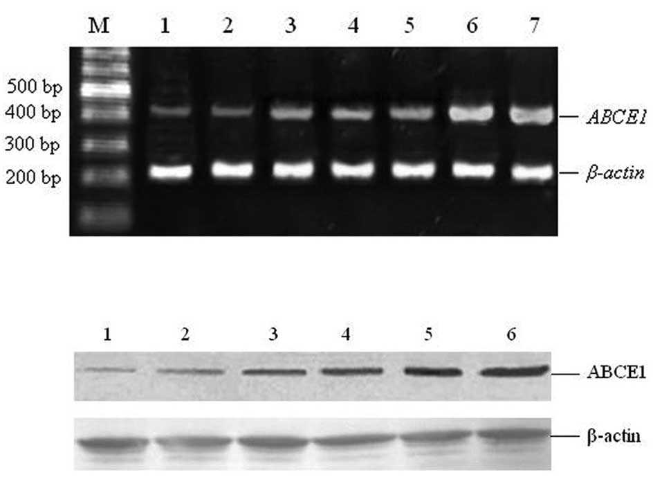
Expression of ABCE1 mRNA and protein in
human lung adenocarcinomas
To probe the role of ABCE1 gene in human lung
adenocarcinoma, we first detected its expression in human lung
adenocarcinoma tissues. The results of RT-PCR and Western blot
analysis showed that ABCE1 mRNA and protein were expressed
in lung adenocarcinoma tissues, normal lung tissues and metastatic
lymph nodes (Fig. 1). By comparing
the expression level in different tissues (17 cases of normal lung
tissues, 18 cases of lung adenocarcinoma tissues and 15 cases of
metastatic lymph nodes), we found that the expression of
ABCE1 mRNA and protein in cancer tissues were higher than in
normal tissues (p<0.05) and lower than in metastatic lymph nodes
(p<0.05) (Tables I and II).
 | Table IRelative expression of ABCE1
mRNA expression in tissues. |
Table I
Relative expression of ABCE1
mRNA expression in tissues.
| Tissues | No. of cases | Relative ABCE1
mRNA expression (densitometry of ABCE1/β-actin) | p-value |
|---|
| Normal lung
tissues | 17 | 0.113±0.003 | |
| Lung adenocarcinoma
tissues | 18 | 0.473±0.05 | <0.05 |
| Metastatic lymph
nodes | 15 | 0.886±0.07 | <0.05 |
| Table IIRelative expression of ABCE1
protein expression in tissues. |
Table II
Relative expression of ABCE1
protein expression in tissues.
| Tissues | No. of cases | Relative ABCE1
protein expression (densitometry of ABCE1/β-actin) | p-value |
|---|
| Normal lung
tissues | 17 | 0.135±0.012 | |
| Lung adenocarcinoma
tissues | 18 | 0.421±0.02 | <0.05 |
| Metastatic lymph
nodes | 15 | 0.84±0.036 | <0.05 |
Subsequently, we analyzed the association between
the clinical stages (classified according to the 1997 TNM
classification of UICC) and ABCE1 expression of the 18 cases
of lung adenocarcinoma. As shown in Tables III and IV, ABCE1 mRNA and protein were
differentially expressed in lung cancers of different clinical
stages. The expression in cases of stage III was significantly
higher than cases of stage I and II. And the expression in group of
N1+2 was significantly higher than cases of N0.
| Table IIIRelative expression of ABCE1
mRNA expression in lung adenocarcinomas of different clinical
stages. |
Table III
Relative expression of ABCE1
mRNA expression in lung adenocarcinomas of different clinical
stages.
| No. of cases | Relative ABCE1
mRNA expression (densitometry of ABCE1/β-actin) | p-value |
|---|
| Stage |
| I | 5 | 0.175±0.021 | |
| II | 8 | 0.282±0.016 | 0.198 |
| III | 5 | 0.551±0.022 | <0.05 |
| Group |
| N0 | 8 | 0.279±0.017 | |
| N1+2 | 10 | 0.784±0.023 | <0.05 |
| Table IVRelative expression of ABCE1
protein expression in lung adenocarcinomas of different clinical
stages. |
Table IV
Relative expression of ABCE1
protein expression in lung adenocarcinomas of different clinical
stages.
| No. of cases | Relative ABCE1
protein expression (densitometry of ABCE1/β-actin) | p-value |
|---|
| Stage |
| I | 5 | 0.193±0.023 | |
| II | 8 | 0.291±0.021 | 0.209 |
| III | 5 | 0.533±0.029 | <0.05 |
| Group |
| N0 | 8 | 0.203±0.018 | |
| N1+2 | 10 | 0.701±0.03 | <0.05 |
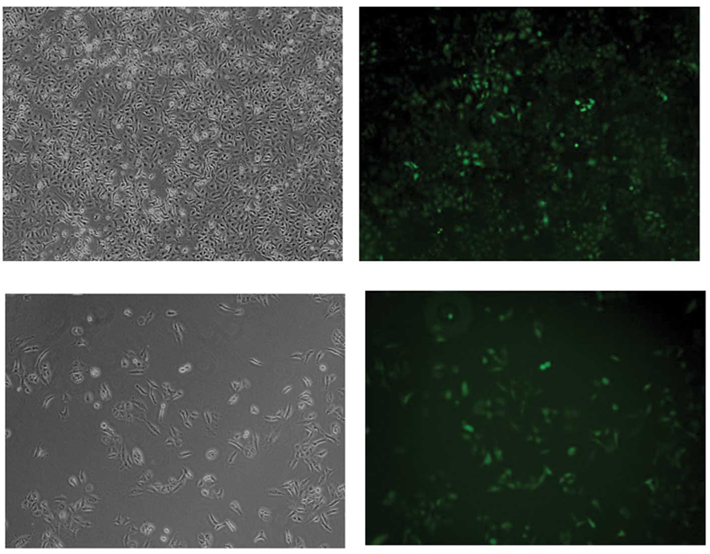
Construction and identification of
lentiviral vectors containing ABCE1-specific shRNA and infection of
human lung adenocarcinoma A549 cells
Specific ABCE1 shRNA fragment was cloned into
the lentiviral pSC-GFP plasmid and confirmed by sequencing (termed
pSC-ABCE1). Then pSC-ABCE1, pCMV-dR8.74 and pMD2G were
co-transfected into incasing 293T cells. Twenty-four hours later,
green fluorescence was detected under a fluorescent microscope
(Fig. 2). Then the supernatant was
collected to obtain lentivirus particles containing pSC-ABCE1. By
infection of 293T cell, the virus titre was calculated as
1×10⁵ IU/ml.
Subsequently, the virus particles were used to
infect human lung adenocarcinoma A549 cells. After 72-h infection,
cells were detected under a fluorescent microscope and the
infection efficiency was >90% by the signals of GFP (Fig. 3).
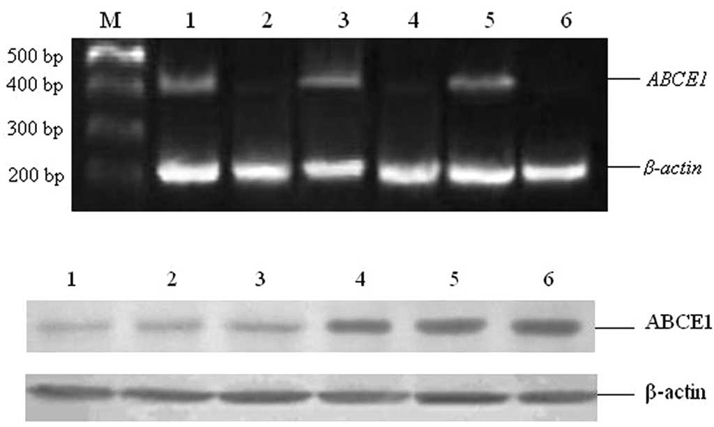
Effect of ABCE1 inhibition on A549 cell
proliferation
To assess the inhibition of ABCE1 expression
after infection, RNA and protein were extracted from control and
lentivirus-infected cells. Then RT-PCR and Western blot analysis
were carried out to detect the ABCE1 mRNA and protein level.
As expected, ABCE1 expression was significantly repressed
after infection (Fig. 4). RT-PCR
showed the densitometry of ABCE1/β-actin was 0.286±0.07 in control
cells, and that in infected cells was 0.023±0.003. The difference
was statistically significant (p<0.05). The results of Western
blot analysis also showed significant difference between the
control and infected cells (the densitometry of ABCE1/β-actin was
0.499±0.097 and 0.109±0.004, respectively p<0.05).
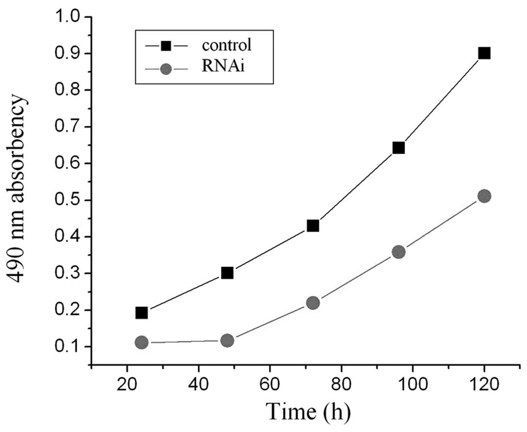
Subsequently, MTT assay was carried out to assess
the proliferation of A549 cells after ABCE1 inhibition.
Cells transfected with or without pSC-ABCE1 were analyzed after 24,
48, 72, 96 or 120-h culture. As shown in Fig. 5, the growth inhibition rate was
42.1, 61.2, 49.1, 44.3 and 43.2%, respectively, with the most
obvious at 48 h, reaching 61.2% and the inhibition was significant
(p<0.05).
Differentially expressed genes after
ABCE1 inhibition
RNA from control or lentivirus-infected A549 cells
was prepared, and then expression chip was used to probe the effect
of ABCE1 inhibition. The results showed 476 evidently
differentially expressed genes (149 genes were up-regulated and 327
ones were down-regulated), including 405 known protein-encoding
genes, which could be divided into the following categories:
inflammatory response factors, signal transduction proteins,
metabolism-related proteins, cell proliferation, development, and
differentiation-related factors, coagulation factors, gene
transcription, translation and modification factors and cell
adhesion and apoptosis-related molecules (Table V).
| Table VPart of the differentially expressed
genes after ABCE1 inhibition. |
Table V
Part of the differentially expressed
genes after ABCE1 inhibition.
| Genes | Description | Expression
change |
|---|
| MTAP | Nucleotide and
nucleic acid metabolism; S-methyl-5-thioadenosine phosphorylase
activity | Up-regulated |
| GADD45 | Activation of MAPKKK
activity; apoptosis; cell differentiation | Up-regulated |
| Caspase-7 | Apoptosis-related
cysteine peptidase | Up-regulated |
| TANK | Signal transduction;
metal ion binding | Up-regulated |
| ZNF14 | Regulation of
transcription, DNA-dependent; zinc ion binding | Up-regulated |
| P27KIP1 | Regulation of cyclin
dependent protein kinase activity, regulation of cell
proliferation | Up-regulated |
| CIDEB | DNA damage response,
signal transduction resulting in induction of apoptosis | Up-regulated |
| TIMP2 | Metalloendopeptidase
inhibitor activity | Down-regulated |
| TNFRSF1B | Cytokine and
chemokine mediated signaling pathway, apoptosis | Down-regulated |
| CDH11 | Homophilic cell
adhesion; calcium ion binding | Down-regulated |
| LAMC1 | Regulation of
epithelial cell proliferation; cell adhesion | Down-regulated |
| TFF3 | Defense response;
digestion | Down-regulated |
| CD55 | Complement
activation, classical pathway; innate immune response | Down-regulated |
| CDK4 | Cell proliferation;
G1/S transition of mitotic cell cycle | Down-regulated |
Discussion
In the present study, we found a high expression
level of ABCE1 mRNA and protein in human lung adenocarcinoma
tissues and metastatic lymph nodes, which was also correlated with
clinical stages. The human lung adenocarcinoma A549 cells were
infected with lentiviral vectors containing ABCE1-specific
shRNA, and resulted in significant inhibition of cell growth.
Subsequently, using microarray assay, a number of differentially
expressed genes were found after ABCE1 suppression. Our
results demonstrated the potential role of ABCE1 in the development
and progress of human lung adenocarcinoma.
It has been reported that mutations of RNase L gene
may be correlated with human tumors. For example, RNase L mutation
and reduction in catalytical activity are found in prostate cancer
and allow tumor cells to escape a potent apoptotic pathway. Due to
the effect of ABCE1 in inhibiting RNase L and its crucial role in
regulating cell growth and proliferation, it is presumed to
participate in the development and progress of human tumors. As
ABCE1 is essential for translation initiation, and translation is a
highly regulated process important to development and pathologies
of cancer, tumor cells are thought to be more sensitive to the
ABCE1 loss of function, making ABCE1 a potential target for
therapeutics (7). In the present
study, to probe the role of ABCE1 in human lung adenocarcinoma, we
first detected its mRNA and protein expression in tissues. By
comparing the expression level in different tissues (normal lung
tissues, lung adenocarcinoma tissues and metastatic lymph nodes),
we found that the expression of ABCE1 mRNA and protein in
cancer tissues were higher than in normal tissues, suggesting a
possible role of ABCE1 in the development of lung adenocarcinoma.
Our study showed, with the advancement of clinical stages of lung
adenocarcinoma, ABCE1 mRNA and protien expression were
increased. The expression in tissues of stage III was significantly
higher than that of stages I–II, and tissues in group N1+2
expressed at higher level of ABCE1 than that in group N0, which
indicated high expression of ABCE1 may possibly induce the growth
and metastasis of lung adenocarcinoma, and detection of ABCE1
expression may help to determine the progress of tumors.
Subsequently, to assess the function of ABCE1 in
lung adenocarcinoma, we carried out siRNA assay. Lentiviral vectors
containing ABCE1-specific shRNA were constructed and human
lung adenocarcinoma A549 cells were infected. The ABCE1 expression
was repressed, cell proliferation was greatly inhibited, with the
most obvious effect at 48 h. Our result was in accordance with the
report that down-regulation of ABCE1 inhibited proliferation of HEK
293 cells (7). These results
demonstrated the role of ABCE1 in cell growth.
To probe the potential function of ABCE1 in lung
adenocarcinoma, we performed microarray analysis to find the
differentially expressed genes after ABCE1 silence. As expected, a
number of genes were found to be up- or down-regulated, which
encoding various types of proteins. The results showed the
expression of 476 genes was evidently changed (149 genes were
up-regulated and 327 ones were down-regulated), including 405 known
protein-encoding genes, which could be divided into categories such
as inflammatory response factors, signal transduction proteins,
metabolism-related proteins, and cell proliferation-related
factors, indicating the wide role of ABCE1 in the physiological and
pathological processes. For example, the expression of GADD45
(growth arrest and DNA damage 45) was increased after ABCE1
silence. As a cell cycle-dependent protein, the expression of
GADD45 is changed following the progress of cell cycle, at the
highest in G1 phase and lower in S phase (8–11). It
participates in maintaining genomic stability, DNA repair and
inhibiting cell growth, which plays a crucial role in the block of
cell transformation and maligant progress. Its abnormal expression
is found in pancreatic cancer, and breast cancer (12,13).
Our study showed silence of ABCE1 gene induced GADD45
expression, and inhibited cell growth. Therefore, it is possible
that ABCE1 repression blocked cells from entry to S phase, which
may result in augmentation of GADD45. However, the accurate
mechanism needs to be further probed.
Collectively, our results demonstrated abnormal
expression of ABCE1 gene in human lung adenocarcinoma and
its effect in the proliferation of lung adenocarcinoma cells. A
number of differentially expressed genes were found after
ABCE1 silencing. The present study may provide some
molecular basis for the mechanisms of development and progress of
human lung adenocarcinoma, and help to find new treatment
targets.
References
|
1
|
Bisbal C, Silhol M, Laubenthal H, et al:
The 2′-5′ oligoadenylate/RNase L/RNase L inhibitor pathway
regulates both MyoD mRNA stability and muscle cell differentiation.
Mol Cell Biol. 20:4959–4969. 2000.
|
|
2
|
Hassel BA, Zhou A, Sotomayor C, Maran A
and Silverman RH: A dominant negative mutant of 2–5A-dependent
RNase suppresses antiproliferative and antiviral effects of
interferon. EMBO J. 12:3297–3304. 1993.
|
|
3
|
Le Roy F, Bisbal C, Silhol M, Martinand C,
Lebleu B and Salehzada T: The 2–5A/RNase L/RNase L inhibitor (RLI)
[correction of (RNI)] pathway regulates mitochondrial mRNAs
stability in interferon alpha-treated H9 cells. J Biol Chem.
276:48473–48482. 2001.
|
|
4
|
Salzberg S, Lanciano F and Hacohen D:
Reversibility of the antiproliferative effect of interferon. Nat
Immun Cell Growth Regul. 9:191–202. 1990.PubMed/NCBI
|
|
5
|
Chen ZQ, Dong J, Ishimura A, et al: The
essential vertebrate ABCE1 protein interacts with eukaryotic
initiation factors. J Biol Chem. 281:7452–7457. 2006. View Article : Google Scholar : PubMed/NCBI
|
|
6
|
Karcher A, Buttner K, Martens B, et al:
X-ray structure of RLI, an essential twin cassette ABC ATPase
involved in ribosome biogenesis and HIV capsid assembly. Structure.
13:649–659. 2005. View Article : Google Scholar : PubMed/NCBI
|
|
7
|
Chen ZQ, Dong J, Ishimura A, Daar I,
Hinnebusch AG and Dean M: The essential vertebrate ABCE1 protein
interacts with eukaryotic initiation factors. J Biol Chem.
28:7452–7457. 2006. View Article : Google Scholar : PubMed/NCBI
|
|
8
|
Fornace AJ Jr, Alamo I Jr and Hollander
MC: DNA damage-inducible transcripts in mammalian cells. Proc Natl
Acad Sci USA. 85:8800–8804. 1988. View Article : Google Scholar : PubMed/NCBI
|
|
9
|
Kastan MB, Zhan Q, el-Deiry WS, et al: A
mammalian cell cycle checkpoint pathway utilizing p53 and GADD45 is
defective in ataxia-telangiectasia. Cell. 71:587–597. 1992.
View Article : Google Scholar : PubMed/NCBI
|
|
10
|
Gujuluva CN, Baek JH, Shin KH, et al:
Effect of UV-irradiation on cell cycle, viability and the
expression of p53, gadd153 and gadd45 genes in normal and
HPV-irnmortalized human oral keratinocytes. Oncogene. 9:1819–1827.
1994.PubMed/NCBI
|
|
11
|
Kearsey JM, Coates PJ, Prescott AR,
Warbrick E and Hall PA: Gadd45 is a nuclear cell cycle regulated
protein which interacts with p21Cip1. Oncogene. 11:1675–1683.
1995.PubMed/NCBI
|
|
12
|
Yamasawa K, Nio Y, Dong M, Yamaguchi K and
Itakura M: Clinicopathological significance of abnormalities in
Gadd45 expression and its relationship to p53 in human pancreatic
cancer. Clin Cancer Res. 8:2563–2569. 2002.PubMed/NCBI
|
|
13
|
Tront JS, Hoffman B and Liebermann DA:
Gadd45a suppresses Ras-driven mammary tumorigenesis by activation
of c-Jun NH2-terminal kinase and p38 stress signaling resulting in
apoptosis and senescence. Cancer Res. 66:8448–8454. 2006.
View Article : Google Scholar : PubMed/NCBI
|