Introduction
Pancreatic cancer has a generally poor prognosis
with a 5-year survival rate of approximately 6% (1). Pancreatic cancer is the 13th most
common cancer and the 4th leading cause of cancer-related deaths in
the world (2–4). Since we do not have an effective
screening method for pancreatic cancer, most patients are diagnosed
at an advanced stage that carries a poor prognosis (2,5). The
discovery of molecular alterations in pancreatic cancer has led to
the development of targeted therapies that have exhibited
substantial benefits in clinical studies, however the survival rate
remains poor (6).
Reactive oxygen species (ROS) consist of radicals,
ions, or other molecules formed by the reduction of oxygen. The
ROS-mediated damage of nucleic acids, proteins and lipids may
affect the process of carcinogenesis (7). ROS may influence the cell cycle
progression, proliferation, cell survival and apoptosis,
angiogenesis and the maintenance of tumor stemness (8). In pancreatic cancer, ROS production is
known to be increased. Contrary to other cancer cells, cancer stem
cells maintain lower intracellular ROS concentrations (9), which may be the result of decreased
ROS production or activated scavenging systems that remove ROS in
cancer stem cells (10,11). Oncogenes that affect different
pathways have been implicated in increases of ROS production. If
oncogenes such as Raf, Myc and cyclin E become overexpressed, ROS
production is increased (12).
Epithelial-mesenchymal transition (EMT) is a crucial process toward
resistance of cell death, chemoresitance, evasion of the immune
system, tumor invasion and tumor metastasis (1,2). In
some authoritative studies, ROS was suggested as mediators or
modulators of the EMT process (3–5).
However, the role of ROS in promoting EMT and cancer stem cell
formation remains unclear.
Ursodeoxycholic acid (UDCA) is a hydrophilic
synthetic bile acid which is the 7-β-epimer of chenodeoxycholic
acid. UDCA is the standard treatment for primary sclerosing
cholangitis, primary biliary cirrhosis, cystic fibrosis and
intrahepatic cholestasis. Although the effect of UDCA as a
therapeutic agent in cancer is uncertain, some studies revealed
such a possibility. Xu et al reported that UDCA had an
anticancer effect in liver cancer cell lines in vitro and in
mice in vivo (13). In
addition, recent studies in murine and rat models revealed that
UDCA had a chemopreventive effect on colorectal cancer (14–17).
UDCA prevented the formation of ROS species and inhibited the Bax
protein-translocation from the cytosol to mitochondria in rat liver
and human hepatocytes. In addition, anti-apoptotic effects were
revealed in other cell types (18–21).
However, the anti-carcinogenic mechanisms of UDCA have not been
elucidated.
UDCA could play important roles via its
anti-apoptotic, anti-inflammatory and chemoprophylactic effects and
for this reason, we investigated the mechanisms of action of UDCA
in pancreatic cancer cells.
Materials and methods
Reagents and materials
UDCA was obtained from Sigma-Aldrich (St. Louis, MO,
USA) and 2′,7′-dichlorofluorescein diacetate (H2DCF-DA) was
purchased from Molecular Probes (Eugene, OR, USA). The antibodies
to sex-determining region Y-box 2 (Sox2) (#4900), E-cadherin
(#3195), N-cadherin (#13116), anti-rabbit IgG (HRP-linked, #7074)
and anti-mouse IgG (HRP-linked, #7076) were purchased from Cell
Signaling Technology (Beverly, MA, USA). Anti-β-actin (LF-PA0207)
and anti-peroxiredoxin II (Prx2) (LF-MA0144) were obtained from
AbFrontier (Seoul, Korea).
Cell lines and culture conditions
The pancreatic cancer cell lines, HPAC and Capan-1
were obtained from Professor Si Young Song (Yonsei University
College of Medicine, Seoul, Korea), cultured in a mixture of
Dulbecco's modified Eagle's medium (DMEM) and F12 containing 10%
fetal bovine serum (FBS) (HPAC), Iscove's modified Dulbecco's
medium (IMDM) containing 20% FBS (Capan-1) with 1%
penicillin-streptomycin, at 37°C in an incubator with a 5%
CO2 atmosphere.
Analysis of ROS by FACS
The level of intracellular ROS was assayed using
H2DCF-DA staining (Thermo Fisher Scientific Inc., Waltham, MA,
USA). To assay the ROS level, 1×10⁶ cells were seeded in
60-mm dishes (Corning Incorporated, Corning, NY, USA). After 12 h,
the seeded cells were treated with 0.2 mM UDCA. After 20 min, the
cells were harvested by trypsin-EDTA and washed with phosphate
buffered saline (PBS). The harvested cells were incubated with 25
µM H2DCF-DA at 37°C for 30 min. The fluorescence intensity was
quantified using flow cytometry (BD Biosciences, Seoul, Korea).
Reverse transcription PCR and
quantitative reverse transcription PCR analyses
Total RNA was extracted from harvested cells using
an RNeasy Plus Mini kit (Qiagen, Hilden, Germany). To remove
genomic DNA, the extracted total RNA was digested by DNase I (New
England BioLabs, Beverly, MA, USA). Purified total RNA in the
amount of 1 µg was reverse transcribed and amplified by RT-PCR
using a High-Capacity cDNA Reverse Transcription kit (Applied
Biosystems, Foster City, CA, USA). The reactions of quantitative
RT-PCR were assessed using SYBR Premix Ex Taq II (Takara, Kusatsu,
Japan) on an iCycler (Bio-Rad Laboratories Inc., Hercules, CA,
USA). The following primer pairs were used: human Prx2 (gene ID:
7001), 5′-TGTGATCGTCCGTGCGTCTA-3′ and 5′-CCGATGCGCGCGTTAC-3′; human
Sox2 (gene ID: 6657), 5′-TGCGAGCGCTGCACAT-3′ and
5′-GCAGCGTGTACTTATCCTTCTTCA-3′; human E-cadherin (gene ID: 999),
5′-CTGAGAACGAGGCTAACG-3′ and 5′-GTCCACCATCATCATTCAATA-3′; human
N-cadherin (gene ID: 1000), 5′-TGGATGGACCTTATGTTGCT-3′ and
5′-AACACCTGTCTTGGGATCAA-3′; GAPDH (gene ID: 2597),
5′-AGGGCTGCTTTTAACTCTGGT-3′ and 5′-CCCCACTTGATTTTGGAGGGA-3′. The
thermal conditions for quantitative reverse transcription PCR assay
were as follows: cycle 1, 95°C for 3 min; cycle 2 (×40), at 95°C
for 10 sec and at 55°C for 30 sec. Target genes were normalized to
GAPDH. The fold change from the untreated control was set at
1-fold, and the normalized fold change ratio was calculated using
the ΔΔCt method.
Immunoblotting
The collected cells were lysed in cold RIPA lysis
buffer (0.5 M Tris-HCl, pH 7.4, 1.5 M NaCl, 2.5% deoxycholic acid,
10% NP-40, 10 mM EDTA) with protease and phosphatase inhibitors
(GenDepot, Barker, CA, USA). The cell lysates were subjected to
SDS-PAGE and transferred onto nitrocellulose membranes (Pall Gelman
Laboratory, Ann Arbor, MI, USA). After being blocked with 8% skim
milk or 5% bovine serum albumin (BSA), the membranes were incubated
with primary antibodies overnight at 4°C. The dilutions used for
each antibody were according to the manufacturer's instructions.
After being washed in PBS with 0.1% Tween, the membranes were
incubated with a secondary antibody conjugated to horseradish
peroxidase. The detection step was performed using WesternBright
ECL HRP substrate (Advansta, Menlo Park, CA, USA).
Floating-sphere formation assay
To assay the formation of HPAC and Capan-1
pancreatic cancer spheres, 4,000 cells/well were seeded in 6-well
ultralow attachment plates (Corning Incorporated) in serum-free
DMEM/F12 (HPAC) or IMDM (Capan-1) with 1X B-27 supplement (50X;
Gibco, Invitrogen, Carlsbad, CA, USA), 10 ng/ml hFGF (#4114TC-01M;
R&D Systems, Minneapolis, MN, USA), 10 ng/ml hEGF (#236-EG-01M;
R&D Systems) and Heparin (Sigma-Aldrich). The plated cells were
incubated at 37°C in a 5% CO2 incubator. Seven days
after seeding, the pancreatic cancer spheres were counted.
Results
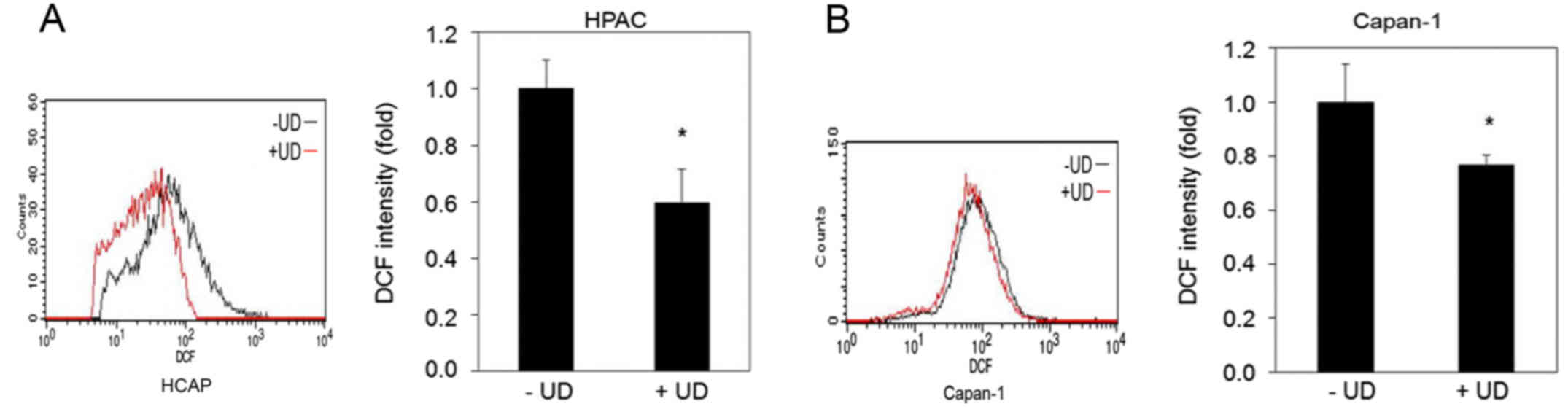
UDCA suppresses the level of ROS in
pancreatic cancer cells through the inhibition of the expression of
Prx2 and a reduction of the phosphorylation of STAT3
To detect the antioxidant effect of UDCA in
pancreatic cancer cells, we assessed the intracellular ROS levels
using DCF-DA staining, as detected by FACS. We found that UDCA
decreased the ROS levels in the HPAC and the Capan-1 cells
(Fig. 1).
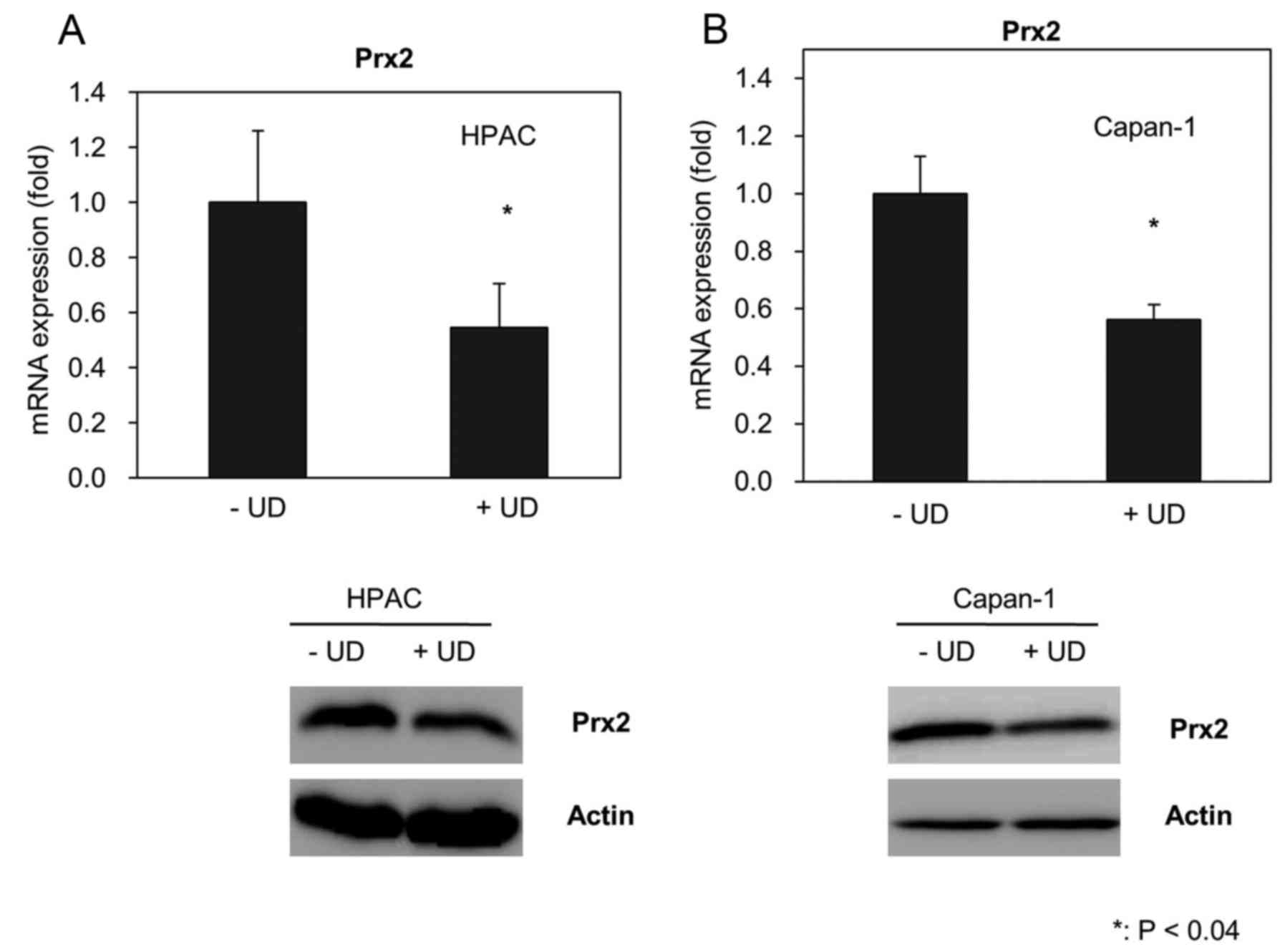
The change of ROS homeostasis by UDCA affected the
expression of antioxidant proteins. Prx2 is an abundant antioxidant
protein in cells. To determine whether UDCA affected the expression
of Prx2, we assessed the level of Prx2 using qRT-PCR and western
blotting. UDCA suppressed the production of Prx2 mRNA in the HPAC
and the Capan-1 cells (Fig. 2).
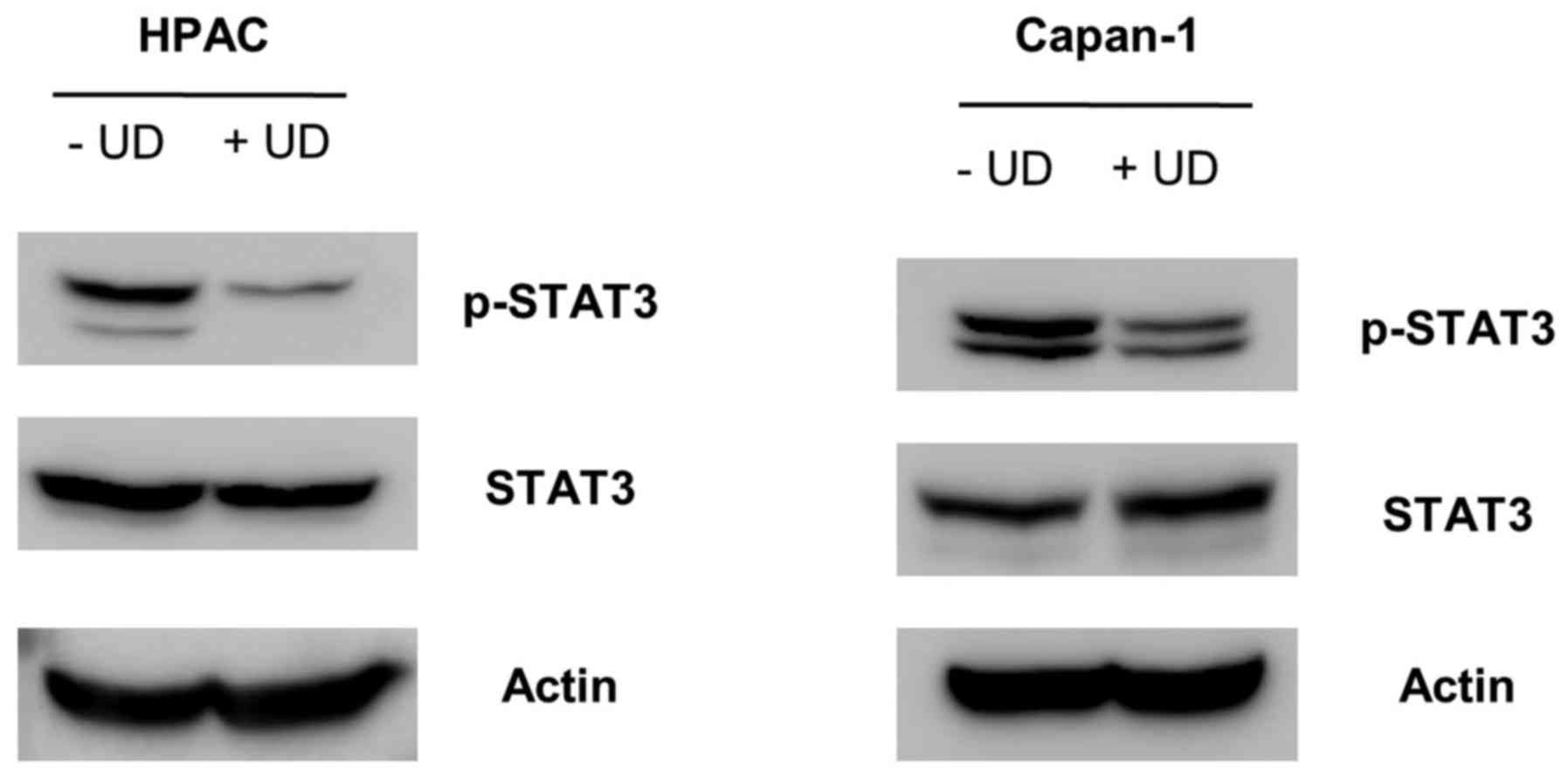
Correspondingly, the protein level of Prx2 was
significantly reduced by UDCA. We observed that treatment of UDCA
inhibited the phosphorylation of STAT3 in the HPAC and the Capan-1
cells (Fig. 3).
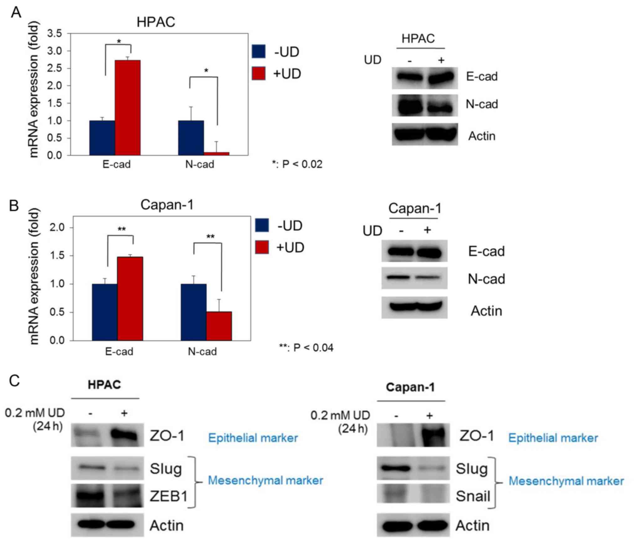
UDCA decreases EMT in pancreatic
cancer cells
UDCA affected the expression of EMT-related
proteins. The expression of E-cadherin mRNA was induced by UDCA in
HPAC cells (Fig. 4A). Furthermore,
the protein level of E-cadherin was increased by UDCA in the HPAC
cells. N-cadherin is a representative marker of mesenchymal cells.
The expression levels of N-cadherin mRNA and protein were
suppressed by UDCA in HPAC cells. The effect of UDCA was replicated
in the Capan-1 cells (Fig. 4B).
UDCA induced the expression of epithelial marker (ZO-1) in the HPAC
and Capan-1 cells. In addition, UDCA suppressed the expression of
Slug and ZEB1 in the HPAC cells. The expression of Snail was
suppressed by UDCA in Capan-1 cells (Fig. 4C).
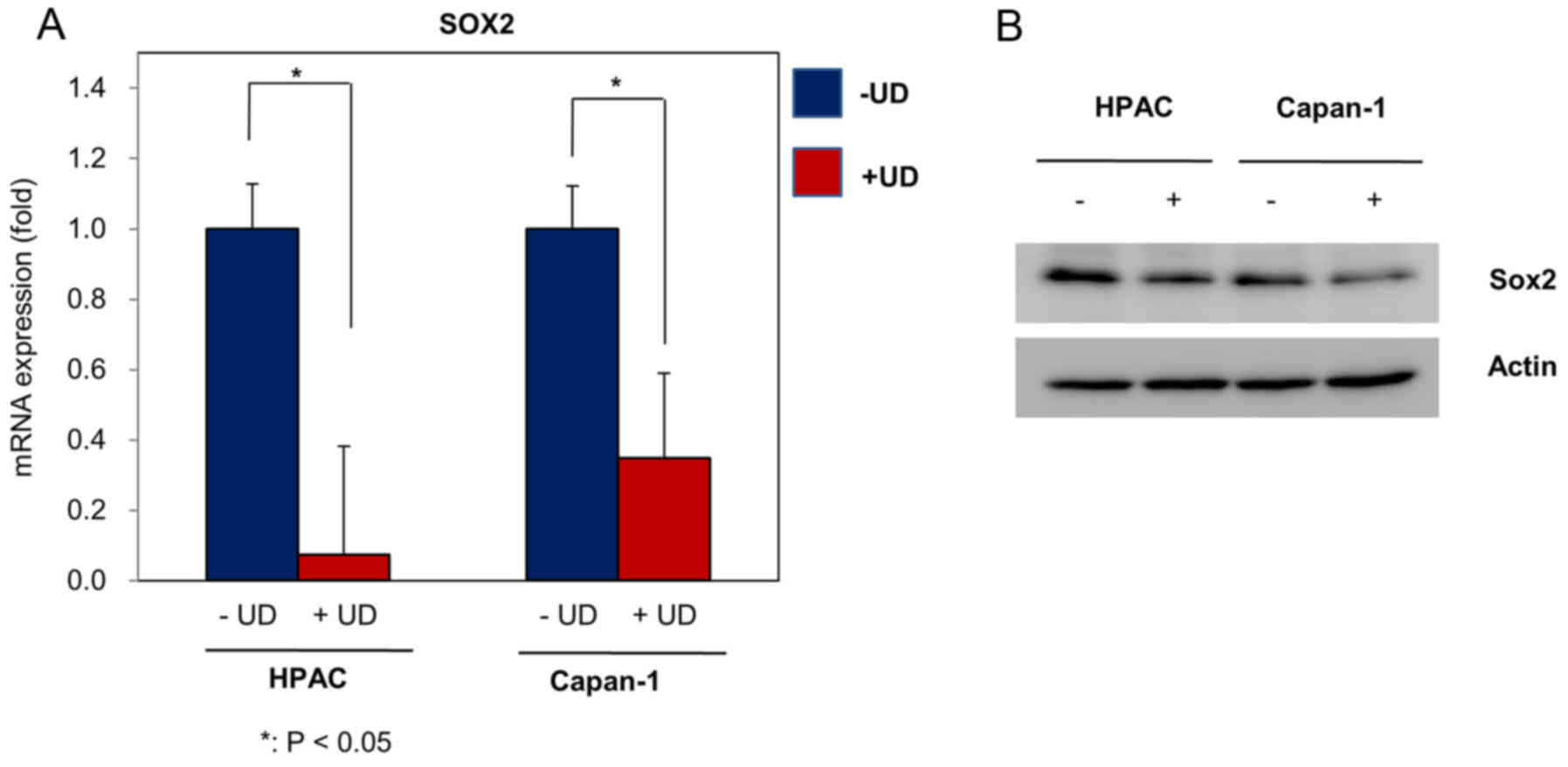
UDCA reduces stemness in pancreatic
cancer cells
The regulation of Sox2 is synchronized with Prx2.
The expression of Sox2 mRNA was suppressed by UDCA in the HPAC and
Capan-1 cells (Fig. 5A). In
addition UDCA reduced the protein level of Sox2 in the HPAC and
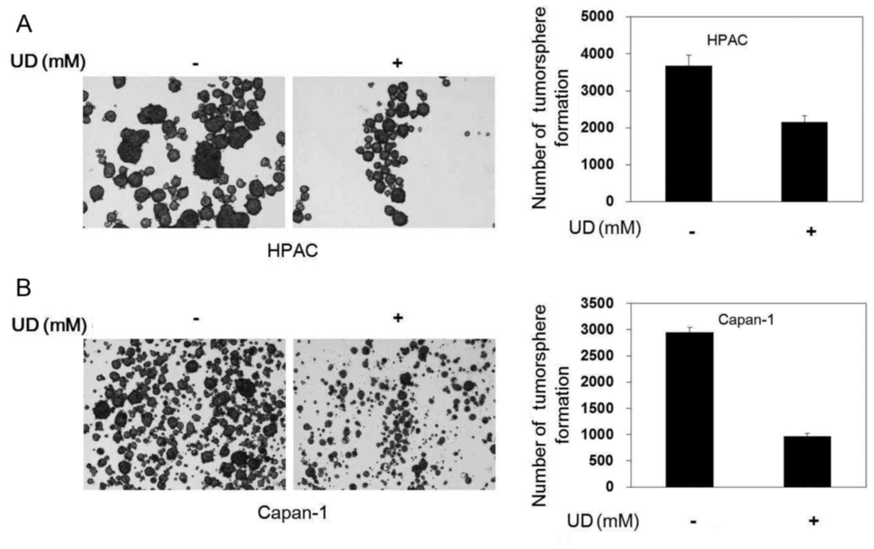
Capan-1 cells (Fig. 5B). Reduced
levels of Sox2 may affect the formation and growth of cancer stem
cells. To investigate whether UDCA reduced the growth of cancer
stem cells, we examined the formation of tumorspheres using culture
in ultralow attachment plates. UDCA reduced the size of
tumorspheres in the HPAC and Capan-1. In addition the total number
of tumorspheres was reduced by UDCA (Fig. 6).
Discussion
UDCA has multifactorial mechanisms of action.
Previous studies (21–24) have revealed that UDCA acts as an
inhibitor of Bax protein translocation from the cytosol to
mitochondria, exhibits a chemopreventive effect on colorectal
cancer, demonstrates anti-apoptotic effects and inhibits the
formation of ROS. UDCA is known as an antioxidant and is a
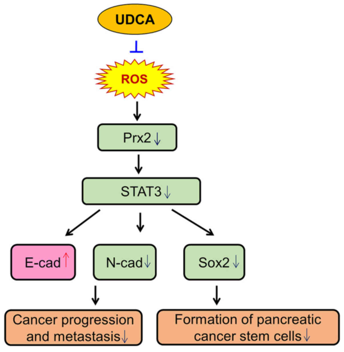
well-tolerated drug (18–20). In the present study, UDCA influenced
the cellular-signaling pathways (Fig.
7). Both in the HPAC and the Capan-1 cells, UDCA did not affect
the same signaling pathway. In the HPAC cells, UDCA did not affect
the phosphrylation of Akt, mTOR, ERK and p38. In the Capan-1 cells,
UDCA reduced the phosphorylation of Akt and mTOR. However, UDCA did
not affect the phosphorylation of ERK and p38 in the Capan-1 cells.
Furthermore the present sudy revealed that treatment with UDCA
reduced the levels of intracellular ROS in the HPAC and Capan-1
cells (Fig. 1).
The lowered level of ROS influenced the cellular
signaling pathways (25,26). The changes in ROS levels can be
related to the concentrations of the antioxidant proteins (25,27–29).
Prx2 is one of the more abundant proteins able to remove ROS in the
cells (27,30). In addition, a recent study revealed
that Prx2 may be associated with drug resistance of cancer stem
cells (31). The present study
revealed that UDCA reduced the mRNA expression and protein level of
Prx2 in pancreatic cancer cells (Fig.
2). Furthemore, UDCA inhibited the expression of Prx1 (data not
shown). The reduced ROS level caused by UDCA did not appear to rely
on the induction of antioxidant proteins.
Prx2 interacts with several molecules in a stressed
environment (27,32). The altered redox state affects the
association between Prx2 and its interacting proteins (33,34).
Prx2 interacts with STAT3 following exposure to
H2O2 (35–37).
This association generates STAT3 oligomerization and increased
phosphorylation of STAT3, which activates the transcriptional
function of STAT3. The present study revealed that UDCA reduced the
amount of phosphorylated STAT3 (Fig.
3). The reduced level of ROS caused by UDCA led to a decrease
in the interaction between Prx2 and STAT3 and suppressed the
phosphorylation of STAT3.
The reduction of STAT3 activation caused by UDCA
affected EMT (38–40). The expression of E-cadherin was
induced by UDCA treatment in the HPAC and Capan-1 cells (Fig. 4). The expression of N-cadherin was
supressed by UDCA in the HPAC and Capan-1 cells (Fig. 4). Thus, the present study revealed
that UDCA suppressed EMT in pancreatic cancer cells. However, the
HPAC and Capan-1 cell lines may not be sufficient to study the EMT
mechanism. Therefore, future experiments using more appropriate
cell lines such as circulating tumor cells or more pancreatic cell
lines as well as cancer patient samples are needed to improve the
study of the EMT mechanism.
Attenuation of the STAT3 activation influenced the
expression of Sox2 (41–43). Recently, a research group revealed
that Prx2 regulated the level of Sox2 (37). As displayed in Fig. 5, the expression of Sox2 was
supressed by UDCA. Sox2 is one of the essential factors in cancer
stem cell pathways. Therefore, the reduction of Sox2 by UDCA
affects the formation of cancer stem cells. In the sphere forming
assay, we found that UDCA reduced the formation of pancreatic
cancer stem cells in the HPAC and Capan-1 cells (Fig. 6). UDCA also suppressed the
maintenance of cancer stem cells in the HPAC and Capan-1 cells
(data not shown).
Pancreatic cancer is a health issue worldwide and is
one of the most aggressive cancers. Due to this fact novel
therapeutic methods are needed for pancreatic cancer, as well as a
more thorough understanding of the genetic and molecular pathways
involved. The present study supports the potential usefulness of
UDCA in pancreatic cancer, due to its antioxidant properties.
However, the anticancer mechanism of UDCA has not been fully
elucidated. Therefore, future studies are warranted to follow up
our results with animal experiments and clinical trials.
In conclusion, UDCA suppressed the level of
intracellular ROS and decreased EMT and stem cell formation in a
pancreatic cancer cell line. Therefore, the antioxidant effects of
UDCA may provide a positive therapeutic benefit for pancreatic
cancer patients.
Acknowledgments
The present study was supported by the Basic Science
Research Program through the National Research Foundation of Korea
(NRF), funded by the Ministry of Education (nos. NRF-2017R1
D1A1B04034547 and NRF-2017R1D1A1B03034546) and Gachon University
Gil Medical Center (2016–15).
References
|
1
|
Knudsen ES, Balaji U, Mannakee B, Vail P,
Eslinger C, Moxom C, Mansour J and Witkiewicz AK: Pancreatic cancer
cell lines as patient-derived avatars: Genetic characterisation and
functional utility. Gut. Jan 10–2017.(Epub ahead of print). doi:
10.1136/gutjnl-2016-313133. View Article : Google Scholar : PubMed/NCBI
|
|
2
|
Stathis A and Moore MJ: Advanced
pancreatic carcinoma: Current treatment and future challenges. Nat
Rev Clin Oncol. 7:163–172. 2010. View Article : Google Scholar : PubMed/NCBI
|
|
3
|
Siegel R, Naishadham D and Jemal A: Cancer
statistics, 2013. CA Cancer J Clin. 63:11–30. 2013. View Article : Google Scholar : PubMed/NCBI
|
|
4
|
Kleger A, Perkhofer L and Seufferlein T:
Smarter drugs emerging in pancreatic cancer therapy. Ann Oncol.
25:1260–1270. 2014. View Article : Google Scholar : PubMed/NCBI
|
|
5
|
Feng W, Zhang B, Cai D and Zou X:
Therapeutic potential of histone deacetylase inhibitors in
pancreatic cancer. Cancer Lett. 347:183–190. 2014. View Article : Google Scholar : PubMed/NCBI
|
|
6
|
Wong HH and Lemoine NR: Pancreatic cancer:
Molecular pathogenesis and new therapeutic targets. Nat Rev
Gastroenterol Hepatol. 6:412–422. 2009. View Article : Google Scholar : PubMed/NCBI
|
|
7
|
Trachootham D, Alexandre J and Huang P:
Targeting cancer cells by ROS-mediated mechanisms: A radical
therapeutic approach? Nat Rev Drug Discov. 8:579–591. 2009.
View Article : Google Scholar : PubMed/NCBI
|
|
8
|
Gupta A, Rosenberger SF and Bowden GT:
Increased ROS levels contribute to elevated transcription factor
and MAP kinase activities in malignantly progressed mouse
keratinocyte cell lines. Carcinogenesis. 20:2063–2073. 1999.
View Article : Google Scholar : PubMed/NCBI
|
|
9
|
Shi X, Zhang Y, Zheng J and Pan J:
Reactive oxygen species in cancer stem cells. Antioxid Redox
Signal. 16:1215–1228. 2012. View Article : Google Scholar : PubMed/NCBI
|
|
10
|
Diehn M, Cho RW, Lobo NA, Kalisky T, Dorie
MJ, Kulp AN, Qian D, Lam JS, Ailles LE, Wong M, et al: Association
of reactive oxygen species levels and radioresistance in cancer
stem cells. Nature. 458:780–783. 2009. View Article : Google Scholar : PubMed/NCBI
|
|
11
|
Ishimoto T, Nagano O, Yae T, Tamada M,
Motohara T, Oshima H, Oshima M, Ikeda T, Asaba R, Yagi H, et al:
CD44 variant regulates redox status in cancer cells by stabilizing
the xCT subunit of system xc− and thereby promotes tumor
growth. Cancer Cell. 19:387–400. 2011. View Article : Google Scholar : PubMed/NCBI
|
|
12
|
Sabharwal SS and Schumacker PT:
Mitochondrial ROS in cancer: Initiators, amplifiers or an Achilles'
heel? Nat Rev Cancer. 14:709–721. 2014. View Article : Google Scholar : PubMed/NCBI
|
|
13
|
Xu Y, Luo Q, Lin T, Zeng Z, Wang G, Zeng
D, Ding R, Sun C, Zhang XK and Chen H: U12, a UDCA derivative, acts
as an anti-hepatoma drug lead and inhibits the mTOR/S6K1 and
cyclin/CDK complex pathways. PLoS One. 9:e1134792014. View Article : Google Scholar : PubMed/NCBI
|
|
14
|
Wali RK, Stoiber D, Nguyen L, Hart J,
Sitrin MD, Brasitus T and Bissonnette M: Ursodeoxycholic acid
inhibits the initiation and postinitiation phases of
azoxymethane-induced colonic tumor development. Cancer Epidemiol
Biomarkers Prev. 11:1316–1321. 2002.PubMed/NCBI
|
|
15
|
Loddenkemper C, Keller S, Hanski ML, Cao
M, Jahreis G, Stein H, Zeitz M and Hanski C: Prevention of
colitis-associated carcinogenesis in a mouse model by diet
supplementation with ursodeoxycholic acid. Int J Cancer.
118:2750–2757. 2006. View Article : Google Scholar : PubMed/NCBI
|
|
16
|
Serfaty L: Chemoprevention of colorectal
cancer with ursodeoxycholic acid: Pro. Clin Res Hepatol
Gastroenterol. 36 Suppl 1:S53–S60. 2012. View Article : Google Scholar : PubMed/NCBI
|
|
17
|
van Heumen BW, Roelofs HM, Morsche Te RH,
Marian B, Nagengast FM and Peters WH: Celecoxib and
tauro-ursodeoxycholic acid co-treatment inhibits cell growth in
familial adenomatous polyposis derived LT97 colon adenoma cells.
Exp Cell Res. 318:819–827. 2012. View Article : Google Scholar : PubMed/NCBI
|
|
18
|
Benz C, Angermüller S, Töx U,
Klöters-Plachky P, Riedel HD, Sauer P, Stremmel W and Stiehl A:
Effect of tauroursodeoxycholic acid on bile-acid-induced apoptosis
and cytolysis in rat hepatocytes. J Hepatol. 28:99–106. 1998.
View Article : Google Scholar : PubMed/NCBI
|
|
19
|
Rodrigues CM, Fan G, Ma X, Kren BT and
Steer CJ: A novel role for ursodeoxycholic acid in inhibiting
apoptosis by modulating mitochondrial membrane perturbation. J Clin
Invest. 101:2790–2799. 1998. View
Article : Google Scholar : PubMed/NCBI
|
|
20
|
Benz C, Angermüller S, Otto G, Sauer P,
Stremmel W and Stiehl A: Effect of tauroursodeoxycholic acid on
bile acid-induced apoptosis in primary human hepatocytes. Eur J
Clin Invest. 30:203–209. 2000. View Article : Google Scholar : PubMed/NCBI
|
|
21
|
Lindor K: Ursodeoxycholic acid for the
treatment of primary biliary cirrhosis. N Engl J Med.
357:1524–1529. 2007. View Article : Google Scholar : PubMed/NCBI
|
|
22
|
Paumgartner G and Beuers U:
Ursodeoxycholic acid in cholestatic liver disease: Mechanisms of
action and therapeutic use revisited. Hepatology. 36:525–531. 2002.
View Article : Google Scholar : PubMed/NCBI
|
|
23
|
Siegel JL, Jorgensen R, Angulo P and
Lindor KD: Treatment with ursodeoxycholic acid is associated with
weight gain in patients with primary biliary cirrhosis. J Clin
Gastroenterol. 37:183–185. 2003. View Article : Google Scholar : PubMed/NCBI
|
|
24
|
Perez MJ and Briz O: Bile-acid-induced
cell injury and protection. World J Gastroenterol. 15:1677–1689.
2009. View Article : Google Scholar : PubMed/NCBI
|
|
25
|
Ray PD, Huang BW and Tsuji Y: Reactive
oxygen species (ROS) homeostasis and redox regulation in cellular
signaling. Cell Signal. 24:981–990. 2012. View Article : Google Scholar : PubMed/NCBI
|
|
26
|
Schieber M and Chandel NS: ROS function in
redox signaling and oxidative stress. Curr Biol. 24:R453–R462.
2014. View Article : Google Scholar : PubMed/NCBI
|
|
27
|
Rhee SG, Woo HA, Kil IS and Bae SH:
Peroxiredoxin functions as a peroxidase and a regulator and sensor
of local peroxides. J Biol Chem. 287:4403–4410. 2012. View Article : Google Scholar : PubMed/NCBI
|
|
28
|
Poljsak B, Šuput D and Milisav I:
Achieving the balance between ROS and antioxidants: When to use the
synthetic antioxidants. Oxid Med Cell Longev. 2013:9567922013.
View Article : Google Scholar : PubMed/NCBI
|
|
29
|
Espinosa-Diez C, Miguel V, Mennerich D,
Kietzmann T, Sánchez-Pérez P, Cadenas S and Lamas S: Antioxidant
responses and cellular adjustments to oxidative stress. Redox Biol.
6:183–197. 2015. View Article : Google Scholar : PubMed/NCBI
|
|
30
|
Wood ZA, Schröder E, Harris Robin J and
Poole LB: Structure, mechanism and regulation of peroxiredoxins.
Trends Biochem Sci. 28:32–40. 2003. View Article : Google Scholar : PubMed/NCBI
|
|
31
|
Cerda MB, Lloyd R, Batalla M, Giannoni F,
Casal M and Policastro L: Silencing peroxiredoxin-2 sensitizes
human colorectal cancer cells to ionizing radiation and
oxaliplatin. Cancer Lett. 388:312–319. 2017. View Article : Google Scholar : PubMed/NCBI
|
|
32
|
Latimer HR and Veal EA: Peroxiredoxins in
regulation of MAPK signalling pathways; sensors and barriers to
signal transduction. Mol Cells. 39:40–45. 2016. View Article : Google Scholar : PubMed/NCBI
|
|
33
|
Moon JC, Hah YS, Kim WY, Jung BG, Jang HH,
Lee JR, Kim SY, Lee YM, Jeon MG, Kim CW, et al: Oxidative
stress-dependent structural and functional switching of a human
2-Cys peroxiredoxin isotype II that enhances HeLa cell resistance
to H2O2-induced cell death. J Biol Chem.
280:28775–28784. 2005. View Article : Google Scholar : PubMed/NCBI
|
|
34
|
Qu D, Rashidian J, Mount MP, Aleyasin H,
Parsanejad M, Lira A, Haque E, Zhang Y, Callaghan S, Daigle M, et
al: Role of Cdk5-mediated phosphorylation of Prx2 in MPTP toxicity
and Parkinson's disease. Neuron. 55:37–52. 2007. View Article : Google Scholar : PubMed/NCBI
|
|
35
|
Park H, Noh AL, Kang JH, Sim JS, Lee DS
and Yim M: Peroxiredoxin II negatively regulates
lipopolysaccharide-induced osteoclast formation and bone loss via
JNK and STAT3. Antioxid Redox Signal. 22:63–77. 2015. View Article : Google Scholar : PubMed/NCBI
|
|
36
|
Sobotta MC, Liou W, Stöcker S, Talwar D,
Oehler M, Ruppert T, Scharf AN and Dick TP: Peroxiredoxin-2 and
STAT3 form a redox relay for H2O2 signaling.
Nat Chem Biol. 11:64–70. 2015. View Article : Google Scholar : PubMed/NCBI
|
|
37
|
Kwon T, Bak Y, Park YH, Jang GB, Nam JS,
Yoo JE, Park YN, Bak IS, Kim JM, Yoon DY, et al: Peroxiredoxin II
is essential for maintaining stemness by redox regulation in liver
cancer cells. Stem Cells. 34:1188–1197. 2016. View Article : Google Scholar : PubMed/NCBI
|
|
38
|
Hawkins K, Mohamet L, Ritson S, Merry CL
and Ward CM: E-cadherin and, in its absence, N-cadherin promotes
Nanog expression in mouse embryonic stem cells via STAT3
phosphorylation. Stem Cells. 30:1842–1851. 2012. View Article : Google Scholar : PubMed/NCBI
|
|
39
|
Xiong H, Hong J, Du W, Lin YW, Ren LL,
Wang YC, Su WY, Wang JL, Cui Y, Wang ZH, et al: Roles of STAT3 and
ZEB1 proteins in E-cadherin down-regulation and human colorectal
cancer epithelial-mesenchymal transition. J Biol Chem.
287:5819–5832. 2012. View Article : Google Scholar : PubMed/NCBI
|
|
40
|
Zhang C, Guo F, Xu G, Ma J and Shao F:
STAT3 cooperates with Twist to mediate epithelial-mesenchymal
transition in human hepatocellular carcinoma cells. Oncol Rep.
33:1872–1882. 2015. View Article : Google Scholar : PubMed/NCBI
|
|
41
|
Foshay KM and Gallicano GI: Regulation of
Sox2 by STAT3 initiates commitment to the neural precursor cell
fate. Stem Cells Dev. 17:269–278. 2008. View Article : Google Scholar : PubMed/NCBI
|
|
42
|
Gao H, Teng C, Huang W, Peng J and Wang C:
SOX2 Promotes the epithelial to mesenchymal transition of
esophageal squamous cells by modulating slug expression through the
activation of STAT3/HIF-α Signaling. Int J Mol Sci. 16:21643–21657.
2015. View Article : Google Scholar : PubMed/NCBI
|
|
43
|
Wang H, Cai HB, Chen LL, Zhao WJ, Li P,
Wang ZQ and Li Z: STAT3 correlates with stem cell-related
transcription factors in cervical cancer. J Huazhong Univ Sci
Technolog Med Sci. 35:891–897. 2015. View Article : Google Scholar : PubMed/NCBI
|