Introduction
Breast cancer (BC) is the most common type of cancer
among females and accounts for ~16% of all cancers (1). BC is a heterogeneous malignant tumor
derived from breast tissue and is the main cause of
cancer-associated mortality in females worldwide (2,3). Surgery
and adjuvant therapy can effectively improve the prognosis of
patients with BC; however, metastasis and recurrence can still
occur (4,5). The development of BC is a complex
process, involving genetic and epigenetic changes (6). Due to the heterogeneity of BC, clinical
outcomes vary. The predictive value of current diagnostic and
prognostic factors for BC is limited. Thus, it is essential to
identify effective diagnostic markers, therapeutic targets and
prognostic factors. Recently, competing endogenous RNAs (ceRNAs)
have been investigated in the process of tumorigenesis and cancer
development (7,8). Salmena et al (7) proposed the ceRNA hypothesis in 2011, in
which a complex post-transcriptional control network involving all
types of RNA transcripts suppresses the functions of microRNAs
(miRNAs/miRs) via miRNA response elements.
Long non-coding RNAs (lncRNAs) are a class of
noncoding RNAs with a length >200 nucleotides (9); a lncRNA may function as an oncogene or a
tumor suppressor to regulate the expression of target genes at
various levels (10,11). Increasing evidence has demonstrated
that lncRNAs serve an important role in several cellular processes
(12,13). The predictive value of lncRNAs has
been reported in multiple types of cancer, including lung cancer
(14), gallbladder cancer (15), ovarian cancer (16), glioblastoma (17), gastric cancer (18), pancreatic cancer (19) and colorectal cancer (20). Abnormal expression of certain lncRNAs
has been observed in BC, and it has been associated with diagnosis
and prognosis of the disease (21).
However, due to the limited number of tissue samples employed, the
results of these studies in BC are inconsistent (22,23).
In the present study, the expression profiles of
mRNAs, miRNAs and lncRNAs in BC and non-cancer tissues were
obtained from The Cancer Genome Atlas (TCGA). An lncRNA-ceRNA
network associated with BC was successfully constructed. In
addition, functional enrichment analysis was conducted to
investigate the potential mechanism. Survival analysis was also
performed for the differentially expressed genes (DEGs) and their
association with BC prognosis was explored. Differentially
expressed RNAs (DERNAs) in the ceRNA network were analyzed to
further investigate the development and prognosis of BC.
Materials and methods
Data acquisition and processing
RNA sequencing (RNA-Seq) and the corresponding
clinical data of BC patients were obtained from the TCGA data
portal (https://tcga-data.nci.nih.gov/tcga/). The lncRNA, mRNA
and miRNA sequence data were derived from Illumina HiSeq RNA Seq
and Illumina HiSeq miRNA Seq platforms. A total of 1,109 BC tissues
and 113 normal breast tissues were included in the present study.
The inclusion criteria were as follows: i) Gender was female; and
ii) histopathology was confirmed as ductal or lobular disease. The
exclusion criteria were as follows: i) Gender was male; and ii 2)
histopathology was not confirmed as ductal and lobular disease. The
present study was conducted in accordance with the publication
guidelines provided by the TCGA (http://cancergenome.nih.gov/publications/publicationguidelines).
Therefore, further approval from an ethics committee was not
required.
Identification of DERNAs
Differentially expressed lncRNA (DElncRNA) and
differentially expressed mRNA (DEmRNA) were defined and encoded
based on the annotations from the Ensembl database (http://www.ensembl.org/index.html). To further
analyze the data, the edgeR package in the R language 3.53 was used
(http://bioconductor.org/packages/edgeR/), and the
DElncRNA, DEmRNA and differentially expressed miRNA (DEmiRNA) were
identified. A |log2 fold change (FC)| >1.5 and false discovery
rate (FDR) adjusted to a P-value <0.01 were set as the
thresholds. Volcano plots and heatmaps of the DERNAs were produced
by using the gplots and heatmap packages in the R platform,
respectively.
Construction of the ceRNA network
The miRcode (http://www.mircode.org/) database (24) was used to predict lncRNA-miRNA
interactions, and the StarBase v2.0 (25) database (http://starbase.sysu.edu.cn/) was used to decode the
miRNA sequences. In addition, miRNA-targeted mRNAs were retrieved
from the miRDB (26), miRTarBase
(27), and TargetScan (28) databases. Only the mRNAs recognized by
all three databases were considered as candidate mRNAs and were
then intersected with the DEmRNAs to determine the DEmRNAs that
were targeted by the DEmiRNAs. Based on the DEmiRNA-DElncRNA and
DEmiRNA-DEmRNA interactions, a coexpression network of DEGs was
constructed, which was visualized using Cytoscape 3.6.1 (29). In addition, the cytoHubba plugin was
used to evaluate the top 15 genes in the network based on
closeness.
Functional enrichment analysis
To understand the underlying biological mechanisms
of the DEmRNAs in the ceRNA crosstalk network, Gene Ontology (GO)
annotation and Kyoto Encyclopedia of Genes and Genomes (KEGG)
pathway analyses were conducted using the DAVID (database for
annotation, visualization, and integrated discovery; http://david.ncifcrf.gov/) online tool (30) and cluster profiler, which is an R
package for functional classification and enrichment of gene
clusters using hypergeometric distribution. The GO plot package of
R software was used to display the results of the GO and KEGG
analyses. GO and KEGG enrichment analyses were based on the
threshold of P<0.05.
Survival analysis
The high expression and low expression groups were
classified according to the median. Survival analysis was performed
for the DERNAs in the ceRNA network using the ‘survival’ package in
R to assess their prognostic value in patients with BC. Survival
curves were generated using the Kaplan-Meier method, and the
log-rank test was used to compare the differences between the
groups. P<0.05 was considered as the threshold for significance
in all analyses.
Results
DEmRNAs, DElncRNAs and DEmiRNAs in
BC
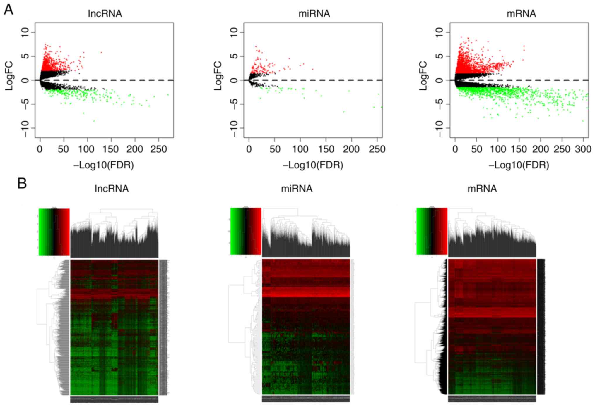
Significant DEGs were identified in 1,109 BC and 113
adjacent non-cancer breast tissues using the ‘edgeR’ package in R
software. A total of 3,198 DEmRNAs (1,996 upregulated and 1,202
downregulated), 1,043 DElncRNAs (814 upregulated and 229
downregulated), and 150 DEmiRNAs (114 upregulated and 36
downregulated) were identified with thresholds of |log2
fold change (FC)| >1.5 and adjusted P<0.01. The distribution
of DEGs based on the two parameters, -log false discovery rate and
logFC, was presented as a volcano plot (Fig. 1A). The heatmap demonstrated the
expression data of all DEGs (Fig.
1B).
Construction of the ceRNA network
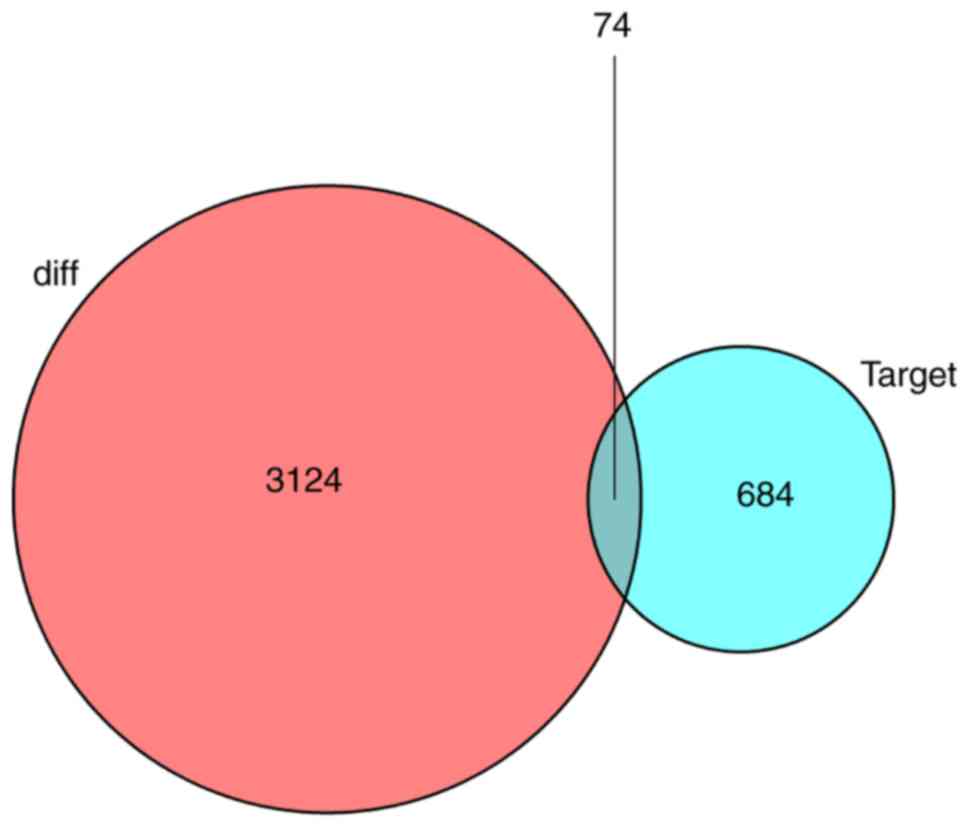
To investigate how lncRNAs mediate mRNA expression
by integrating with miRNAs in BC, a lncRNA-miRNA-mRNA (ceRNA)
network was generated and visualized using Cytoscape v3.6.1. A
total of 405 pairs of interacting lncRNAs and miRNAs were
identified using the Perl program 5.30.0 (http://www.perl.org/) (31), based on the 1,043 DElncRNAs. Based on
24 miRNAs, target mRNAs were identified using the miRTarBase, miRDB
and TargetScan databases. Target genes that were included in all
three datasets were selected. Finally, there were 74 DEmRNAs in the
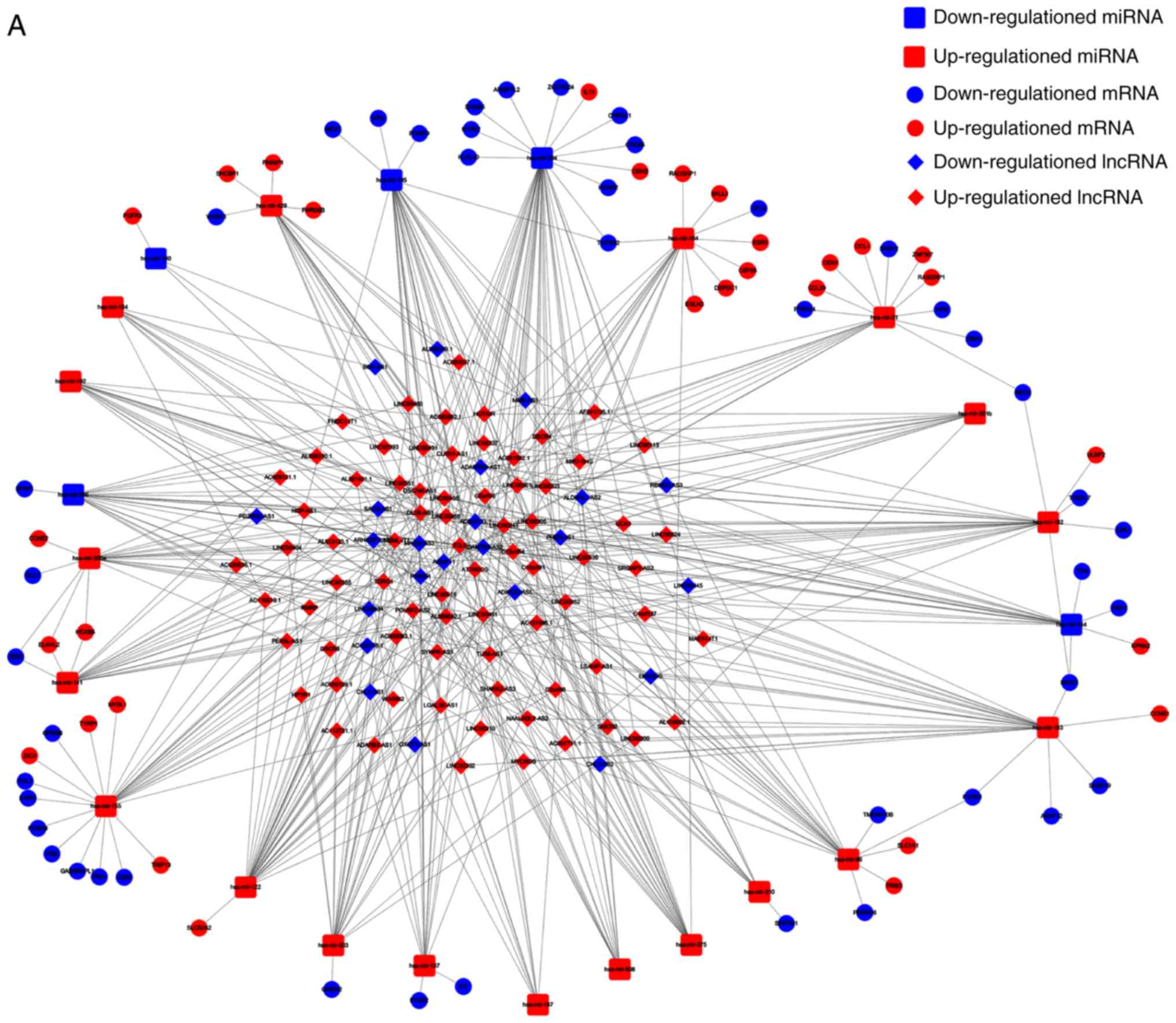
ceRNA network (Fig. 2). In total, 97
lncRNA nodes, 24 miRNA nodes and 74 mRNA nodes were identified as
having differential expression profiles in the ceRNA network
(Fig. 3A). Based on the plug-in
cytoHubba in Cytoscape, the hub network was obtained through the
existing network analysis and closeness scores (Fig. 3B). The results included six miRNAs,
namely hsa-miR-204, hsa-miR-145, hsa-miR-122, hsa-miR-155,
hsa-miR-182 and hsa-miR-183. The lncRNAs involved included
LINC00461, AGAP11, LINC00466, LINC00261, MAGI2-AS3, TCL6,
ADAMTS9-AS2, AL589642.1 and DLX6-AS1.
Functional analysis of DEmRNAs in the
ceRNA network
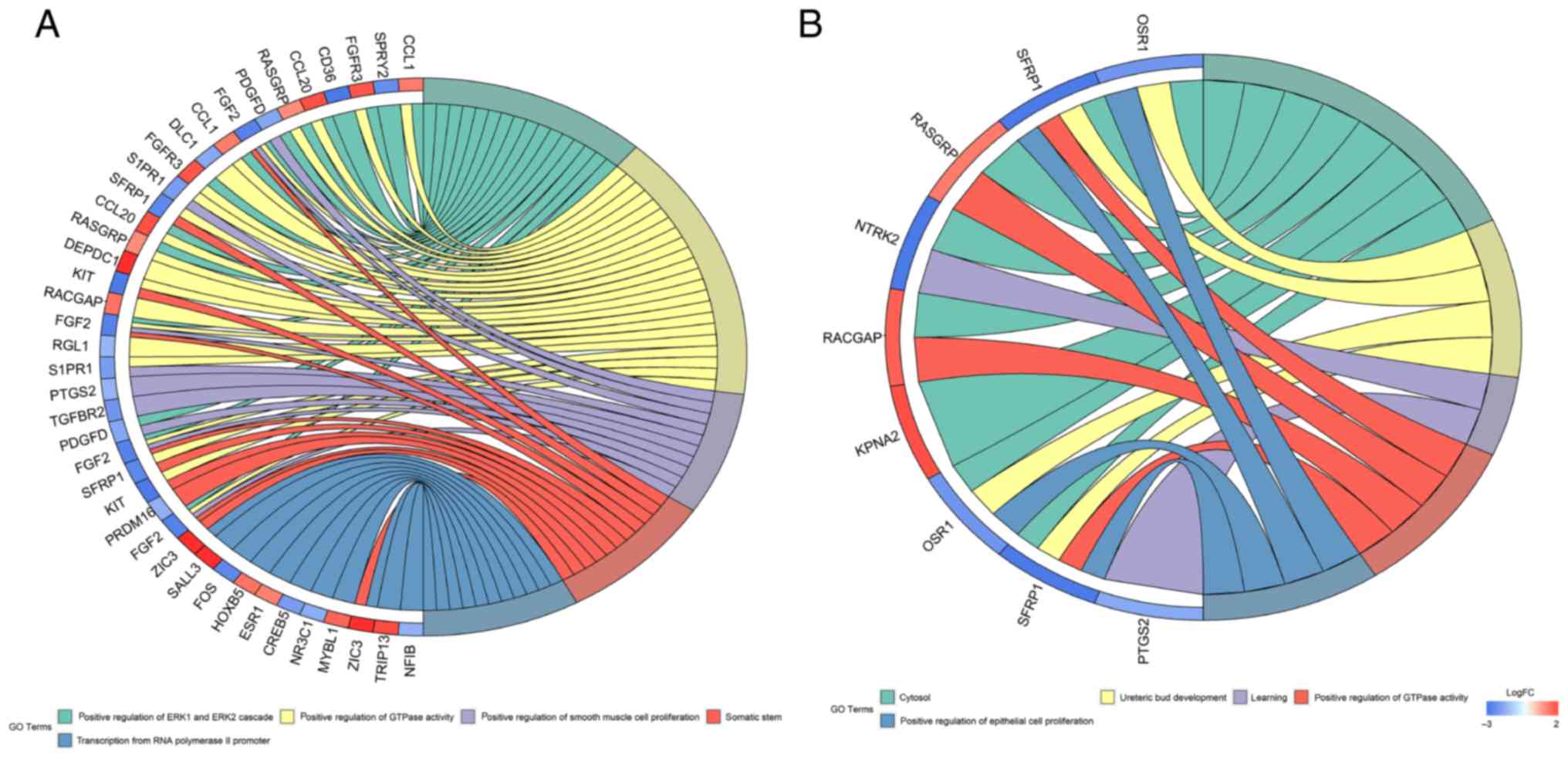
Using GO and KEGG analysis, the biological functions
of the 74 DEmRNAs were investigated. The results indicated that the
DEmRNAs were enriched in two GO biological process categories
(P<0.05; Fig. 4A). The main
biological process GO term was ‘positive regulation of GTPase
activity’. In addition, enriched GO terms based on the analysis of
survival curves associated with KM-significant DEmRNAs were
identified (Fig. 4B). The network
included nine DEmRNAs, which were mainly enriched in ‘positive
regulation of epithelial cell proliferation’.
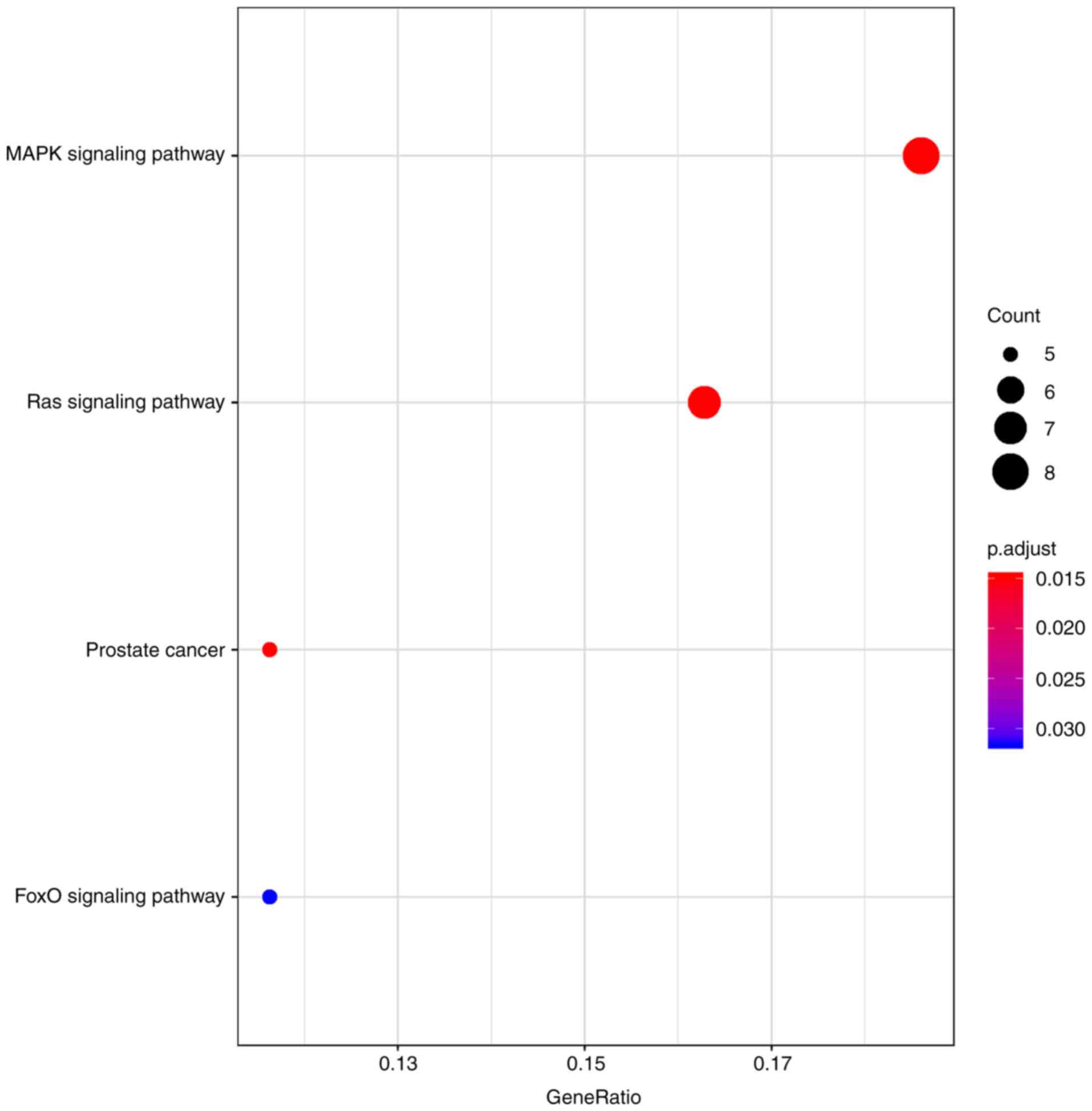
From KEGG analysis, four main enriched pathways were
determined (Table I and Fig. 5), including the ‘mitogen-activated
protein kinase (MAPK) signaling pathway’, the ‘Ras signaling
pathway’, ‘prostate cancer’ and the ‘FoxO signaling pathway’. Among
the four pathways, the ‘MAPK pathway’ and the ‘Ras pathway’ are
associated with BC progression (3,30). In
addition, ‘prostate cancer’ is linked to tumor-related pathways
(32), whereas the ‘FoxO signaling
pathway’ is associated with apoptosis, cell-cycle control and
glucose metabolism (33).
 | Table I.Kyoto Encyclopedia of Genes and
Genomes pathway enrichment analysis of the differentially enriched
mRNAs. |
Table I.
Kyoto Encyclopedia of Genes and
Genomes pathway enrichment analysis of the differentially enriched
mRNAs.
| Pathway ID | Description | Genes involved | Count | P-value | Adjusted P-value
(FDR) |
|---|
| hsa04010 | MAPK signaling
pathway | TGFBR2, KIT, FGF2,
PDGFD, FOS, NTRK2, FGFR3, RASGRP1 | 8 | <0.001 | 0.014 |
| hsa05215 | Prostate
cancer | FOXO1, ERG, CREB5,
PDGFD, CCNE2 | 5 | <0.001 | 0.014 |
| hsa04014 | Ras signaling
pathway | KIT, FGF2, RGL1,
PDGFD, NTRK2, FGFR3, RASGRP1 | 7 | <0.001 | 0.014 |
| hsa04068 | FoxO signaling
pathway | TGFBR2, FOXO1,
GABARAPL1, S1PR1, CCNB1 | 5 | <0.001 | 0.032 |
Survival-associated DERNAs in the
ceRNA network
KM analysis was performed to investigate the
association between DERNAs (DElncRNAs, DEmRNAs and DEmiRNAs) in the
ceRNA network and the prognosis of patients with BC, thereby
obtaining a prognostic signature for each DERNA.
In total, 6 out of the total 97 DElncRNAs were
significantly associated with the rate of overall survival. Among
them, three DElncRNAs (INC AC112721.1, LINC00536 and MIR7-3HG) were
negatively associated with OS, while the other three (ADAMTS9-AS1,
AL356479.1 and LINC00466) were positively associated with OS
(Fig. 6A).
In addition, 9 out of the total 74 DEmRNAs were
significantly associated with the rate of overall survival. Among
them, four DEmRNAs [importin subunit α 2 (KPNA2), Rac
GTPase-activating protein 1 (RACGAP1), Shc binding and spindle
associated 1 (SHCBP1) and zinc finger protein 367 (ZNF367)] were
negatively associated with OS, while the remaining five DEmRNAs
[tropomyosin receptor kinase B (NTRK2), ORS1,
prostaglandin-endoperoxidase synthase 2 (PTGS2), RAS
guanyl-releasing protein 1 (RASGRP1) and secreted frizzled-related
protein 1 (SFRP1)] were positively associated with OS (Fig. 6B).
Finally, 2 out of the total 24 DEmiRNAs were
significantly associated with the rate of OS. The results
demonstrated that hsa-miR-301b was negatively associated with OS,
while hsa-miR-204 was positively associated with OS (Fig. 6C).
Discussion
BC is a common gynecological malignancy; the
incidence of this disease is increasing (3). Despite the combined use of surgery,
chemotherapy, radiotherapy and endocrine therapy, BC remains a
major cause of cancer-associated mortality in females (34). To improve the clinical status of
patients with BC, and develop additional methods for the diagnosis,
treatment and prognosis of this disease, it is crucial to
understand the regulatory mechanism underlying its occurrence and
development. The study of prognostic markers associated with BC may
aid early detection and individualized treatment.
Recently, research into the lncRNA-miRNA-mRNA
network has attracted increasing attention. Non-coding (nc) RNAs
include various forms of RNA, which are categorized into lncRNAs
and short ncRNAs. As short-chain ncRNAs, miRNAs are ~22 nucleotides
in length and bind to sequences with partial complementarity to RNA
transcripts to act as negative regulators of target gene expression
(35). lncRNAs are defined as
transcripts >200 nucleotides in length with no protein-coding
functions (36). Denzler et al
(37) proposed the ‘competitive
endogenous RNA’ hypothesis, suggesting that ceRNAs could regulate
miRNAs. Salmena et al (7)
reported that all types of RNAs can compete with each other for
miRNAs, resulting in large-scale transcription crosstalk throughout
the transcriptome.
Investigation into ceRNAs and ceRNA networks may
provide insight into the processes underlying certain diseases and
aid the development of novel therapeutic strategies. The imbalance
in lncRNA expression is associated with tumorigenesis, metastasis
and prognosis of various types of cancer (14,16,20,38),
suggesting that lncRNAs may be potential biomarkers of cancer. In
order to evaluate the systematic biological function of the
DElncRNAs in the ceRNA network, the present study investigated the
association between DElncRNAs and OS. To comprehensively understand
how the lncRNA-associated ceRNA network affects BC, large-scale
sequencing data of patients with BC were obtained for analysis from
TCGA. DERNAs were detected and a lncRNA-miRNA-mRNA network was
constructed based on the prediction of associated tumor-specific
biological processes. In total, 97 lncRNA, 22 miRNA and 74 mRNA
nodes in the ceRNA networks were identified to be differentially
expressed. In addition, the association between DElncRNAs and OS,
and the biological role of DElncRNAs in ceRNA networks were
investigated. In the present study, nine DEmRNAs (KPNA2, NTRK2,
ORS1, PTGS2, RACGAP1, RASGRP1, SFRP1, SHCBP1 and ZNF367), six
DElncRNAs (INC AC112721.1, ADAMTS9-AS1, AL356479.1, LINC00466,
LINC00536 and MIR7-3HG), and two DEmiRNAs (hsa-miR-204,
hsa-miR-301b) were associated with OS; thus, these DERNAs may serve
as potential prognostic biomarkers of OS in patients with BC.
The regulation of miRNAs is a critical process
associated with tumor-related pathways involved in oncogenesis and
tumor progression (39). It has been
reported that a higher grade of clear cell renal cell carcinoma is
correlated with a concomitant decrease in miR-204 expression
(40,41). Ying et al (42) revealed that miR-204 suppresses the
stem cell-associated phenotype, and the self-renewal and migration
potentials of glioma cells by targeting SRY-box transcription
factor 4 and the EPH receptor B2. Thus, loss of miR-204 expression
enhances tumor migration. Shen et al (43) suggested that miR-204 regulates the
biological behavior of BC cells by directly targeting forkhead box
A1, acting as a tumor suppressor in BC. These reports are
consistent with the current results, in which the increased
expression of miR-204 was associated with improved overall survival
in patients with BC. The current results also demonstrated that
upregulated hsa-miR-301b was associated with poor survival in
patients with BC. Fort et al (44) revealed that the miR-130b/301b cluster
serves a protumorigenic role in prostate cancer.
In the present study, increased expression of
ADAMTS9-AS1, AL356479.1 and LINC00466 was positively associated
with prognosis, while the reduced expression of the remaining three
lncRNAs (INC AC112721.1, LINC00536 and MIR7-3HG) was associated
with improved prognosis. As an antisense lncRNA, ADAMTS9-AS1 serves
crucial roles in the progression and prognosis of carcinoma
(45). ADAMTS9-AS1 was determined to
be associated with the prognosis of patients with bladder cancer
(46,47). Xing et al (48) suggested that ADAMTS9-AS1 may be a
marker for predicting the prognosis of colorectal carcinoma. Li
et al (49) reported that
ADAMTS9-AS1 may be indispensable in the development of the ectoderm
and epithelial cells, and can be effectively employed for
determining the risk of esophageal cancer development. In addition,
it was indicated that ADAMTS9-AS1 serves as a risk-predicting
lncRNA in ovarian cancer (50).
Analysis of BC data revealed ADAMTS9-AS1 and LINC00536 to be
prognostic markers for BC (50).
These previous findings were consistent with the results of the
present study. To the best of our knowledge, no studies have
reported the roles of AC112721.1, AL356479.1, LINC00466 and
MIR7-3HG in cancer.
Accumulating evidence has demonstrated that miRNAs
serve an important role in cancer. In the ceRNA network, the
present study reported that the upregulated expression of LINC00466
was associated with downregulation of hsa-miR-204 and hsa-miR-144;
and MIR7-3HG was determined to downregulate hsa-miR-204 and
hsa-miR-145. Several studies have reported that the levels of
hsa-miR-204 were decreased in certain types of cancer, including
gastric cancer (51) and lung cancer
(52). Based on TCGA data of 1,077 BC
tissue samples, Alhawaj et al (53) determined that miR-204-5p was markedly
downregulated when compared with 104 normal tissue samples. Liu
et al (54) demonstrated that
trichostatin A and tamoxifen inhibited BC cell growth via miR-204
and estrogen receptor (ER) α-mediated suppression of the AKT/mTOR
pathway, suggesting that miR-204 may be associated with tumor
growth and with sensitivity to chemotherapeutic agents. The present
study proposed the involvement of the LINC00466-hsa-miR-204-NTRK2
axis in BC. Thus, LINC00466 and MIR7-3HG may be crucial for the
progression of tumors by interacting with hsa-miR-204. In addition,
hsa-miR-144 and hsa-miR-145 exhibit inverse expression profiles in
BC tissues (55–57).
Combining the results of the hub networks based on
analysis with the plug-in cytoHubba in Cytoscape, the present study
determined that hsa-miR-155 and AGAP11 had a high degree of
closeness, and exhibited significance from GO analysis. Hsa-miR-155
is a multifunctional small RNA located in chromosome 21, and is
involved in a variety of biological and pathological processes
(58). Volinia et al (59) reported that miRNAs served complex
roles in tumors; network analysis indicated that the majority of
miRNAs were linked to hsa-miR-155 within transgenic mice with
miR-155-induced acute lymphocytic leukemia (59). Chuang et al (60) identified that the increased expression
of hsa-miR-155-5p was a poor prognostic marker in acute myeloid
leukemia. Braun et al (61)
revealed that germline polymorphisms at AGAP11 were associated with
stage-adjusted survival in ovarian cancer. In the current network,
hsa-miR-155 was highly expressed, while AGAP11 was expressed at low
levels, and may be a prognostic marker in BC. Additionally, 74
DEmRNAs were identified in the current ceRNA network. GO analysis
revealed that the main enriched GO biological processes were
‘positive regulation of GTPase activity’, ‘positive regulation of
ERK1 and ERK2 cascade’ and ‘transcription from RNA polymerase II
promoter’. Analysis of the enriched GO networks of the
KM-significant DEmRNAs revealed that nine DEmRNAs were mainly
enriched in ‘cytosol’ and ‘positive regulation of epithelial cell
proliferation’. Among the nine DEmRNAs, upregulated RASGRP1
expression was demonstrated to be associated with favorable outcome
in BC. As a guanine nucleotide exchange factor, RASGRP1 can
sensitize Ras and ERK/MAPK cascades. Alterations in RASGRP1
expression have been associated with a variety of diseases,
including systemic lupus erythematosus (62), hepatotoxicity (63), and cancer (64,65).
Upregulated RASGRP1 expression correlates with improved clinical
outcomes in patients with colorectal cancer, and may induce a
negative feedback loop to suppress proliferative epidermal growth
factor receptor/son of sevenless homolog 1/Ras signaling (64). RASGRP1 may be a novel therapeutic
target for the treatment of hepatocellular carcinoma (66). Wang et al (67) reported a positive correlation between
RASGRP1 overexpression and survival in triple-negative breast
cancer. Some studies have reported that RASGRP1 is associated with
the acquired drug resistance of tumors (68,69). In
addition, seven other genes identified in the present study (KPNA2,
NTRK2, PTGS2, RACGAP1, SFRP1, SHCBP1 ZNF367) have been reported to
be associated with the prognosis of tumors (70–75).
Pathway analysis suggested that DEGs were mainly
involved in the ‘MAPK signaling pathway’ and the ‘Ras signaling
pathway’ in the present study. Additionally, the present results
demonstrated that RASGRP1 was enriched in the ‘MAPK signaling
pathway’ and the ‘Ras signaling pathway’. MAPKs are a class of
serine/threonine protein kinases in cells, and the MAPK signaling
pathway is present in most types of cells. MAPK signaling serves an
important role in transducing extracellular signals into cells and
their nuclei, and induces various cellular biological processes,
including cell differentiation, proliferation and apoptosis.
Numerous studies have demonstrated that the MAPK signaling pathway
is associated with the development of BC (76–79). Yang
et al (78) indicated that C-C
motif chemokine ligand 28 has been associated with the
proliferation and metastasis of BC via the MAPK-mediated
anti-apoptotic and metastasis signaling pathways. The androgen
receptor pathway is a potential target for the treatment of BC, and
has been reported to exhibit crosstalk with the MAPK pathway
(77). Previously, the MAPK signaling
pathway was demonstrated to be involved in the development of drug
resistance in tumors (78). Jia et
al (79) reported that
Kruppel-like factor 4 (KLF4) could enhance the response of BC cells
to tamoxifen by inhibiting the MAPK signaling pathway, indicating
that targeting KLF4/MAPK signaling may be a potential therapeutic
strategy for treating BC. RAS is one of the most commonly mutated
genes linked to human cancer. Anticancer inhibitors targeting the
RAS signaling pathway are potential therapeutic agents for the
treatment of cancers; however, further investigation is required.
The abnormal activation of the Ras/ERK pathway mediates the
occurrence and invasiveness of BC, which is notably common in BC.
Ras activation is a crucial determinant of the transmission and
poor prognosis of ERα+/luminal BC (80,81).
To the best of our knowledge, the present study is
the first to report the aberrant expression of INC AC112721.1,
AL356479.1, LINC00466 and MIR7-3HG in BC, indicating their
potential prognostic value in BC. The present study proposed that
LINC00466 and MIR7-3HG interact with hsa-miR-204, which may be
involved in the development of BC; to the best of our knowledge,
this has not been reported previously. Of note, further
bioinformatics analysis of the ceRNA network in BC may aid future
investigations. The current results determined that the genes
associated with survival in BC were enriched in the ‘MAPK signaling
pathway’, which has been reported to be associated with drug
resistance. Our group aims to further investigate the pathology of
BC via in vivo and in vitro experiments in the
future.
Acknowledgements
Not applicable.
Funding
This work was supported by the Science and Education
for Health Foundation of Suzhou for Youth (grant nos. kjxw2018030
and kjxw2018032), the Science and Technology Project Foundation of
Suzhou (grant no. SS201651), the Education Research Project
Foundation of Nanjing Medical University (grant no. FZS-ZD-201701)
and the Key Medical Disciplines in Jiangsu Province (grant no.
ZDXKC2016007).
Availability of data and materials
The datasets used and/or analyzed during the present
study are available from the corresponding author on reasonable
request.
Authors' contributions
JJW, YQH and WS carried out data acquisition and
processing, and were the major contributors in writing the
manuscript. YFL and HW made contributions to data analysis,
interpretation and figure editing. WJW and MH directed the whole
study, and revised the manuscript. All authors read and approved
the final manuscript.
Ethics approval and consent to
participate
Not applicable.
Patient consent for publication
Not applicable.
Competing interests
The authors declare that they have no competing
interests.
References
|
1
|
Siegel R, Ma J, Zou Z and Jemal A: Cancer
statistics, 2014. CA Cancer J Clin. 64:9–29. 2014. View Article : Google Scholar : PubMed/NCBI
|
|
2
|
Wang Y, Zhou Y, Yang Z, Chen B, Huang W,
Liu Y and Zhang Y: MiR-204/ZEB2 axis functions as key mediator for
MALAT1-induced epithelial-mesenchymal transition in breast cancer.
Tumour Biol. 39:10104283176909982017. View Article : Google Scholar : PubMed/NCBI
|
|
3
|
Kroenke CH, Michael YL, Poole EM, Kwan ML,
Nechuta S, Leas E, Caan BJ, Pierce J, Shu XO, Zheng Y and Chen WY:
Postdiagnosis social networks and breast cancer mortality in the
after breast cancer pooling project. Cancer. 123:1228–1237. 2017.
View Article : Google Scholar : PubMed/NCBI
|
|
4
|
Luschin G and Habersack M: Oral
information about side effects of endocrine therapy for early
breast cancer patients at initial consultation and first follow-up
visit: An online survey. Health Commun. 29:421–426. 2014.
View Article : Google Scholar : PubMed/NCBI
|
|
5
|
Cheng Y, Tao L, Xu J, Li Q, Yu J, Jin Y,
Chen Q, Xu Z, Zou Q and Liu X: CD44/cellular prion protein interact
in multidrug resistant breast cancer cells and correlate with
responses to neoadjuvant chemotherapy in breast cancer patients.
Mol Carcinog. 53:686–697. 2014. View
Article : Google Scholar : PubMed/NCBI
|
|
6
|
Bao L, Messer K, Schwab R, Harismendy O,
Pu M, Crain B, Yost S, Frazer KA, Rana B, Hasteh F, et al:
Mutational profiling can establish clonal or independent origin in
synchronous bilateral breast and other tumors. PLoS One.
10:e01424872015. View Article : Google Scholar : PubMed/NCBI
|
|
7
|
Salmena L, Poliseno L, Tay Y, Kats L and
Pandolfi PP: A ceRNA hypothesis: The Rosetta Stone of a hidden RNA
language? Cell. 146:353–358. 2011. View Article : Google Scholar : PubMed/NCBI
|
|
8
|
Rapicavoli NA, Qu K, Zhang J, Mikhail M,
Laberge RM and Chang HY: A mammalian pseudogene lncRNA at the
interface of inflammation and anti-inflammatory therapeutics.
Elife. 2:e007622013. View Article : Google Scholar : PubMed/NCBI
|
|
9
|
Mercer TR, Dinger ME and Mattick JS: Long
non-coding RNAs: Insights into functions. Nat Rev Genet.
10:155–159. 2009. View
Article : Google Scholar : PubMed/NCBI
|
|
10
|
Ponting CP, Oliver PL and Reik W:
Evolution and functions of long noncoding RNAs. Cell. 136:629–641.
2009. View Article : Google Scholar : PubMed/NCBI
|
|
11
|
Anamaria N, Soumillon M, Warnefors M,
Liechti A, Daish T, Zeller U, Baker JC, Grützner F and Kaessmann H:
The evolution of lncRNA repertoires and expression patterns in
tetrapods. Nature. 505:635–640. 2014. View Article : Google Scholar : PubMed/NCBI
|
|
12
|
Xue B and He L: An expanding universe of
the non-coding genome in cancer biology. Carcinogenesis.
35:1209–1216. 2014. View Article : Google Scholar : PubMed/NCBI
|
|
13
|
Xie C, Yuan J, Li H, Li M, Zhao G, Bu D,
Zhu W, Wu W, Chen R and Zhao Y: NONCODEv4: Exploring the world of
long non-coding RNA genes. Nucleic Acids Res. 42((Database Issue)):
D98–D103. 2014. View Article : Google Scholar : PubMed/NCBI
|
|
14
|
Kumar M.S..Armenteros-Monterroso E, East
P, Chakravorty P, Matthews N, Winslow MM and Downward J: HMGA2
functions as a competing endogenous RNA to promote lung cancer
progression. Nature. 505:212–217. 2014. View Article : Google Scholar : PubMed/NCBI
|
|
15
|
Wang C, Chen L, Yang Y, Zhang M and Wong
G: Identification of bladder cancer prognostic biomarkers using an
ageing gene-related competitive endogenous RNA network. Oncotarget.
8:111742–111753. 2017. View Article : Google Scholar : PubMed/NCBI
|
|
16
|
Zhou M, Wang X, Shi H, Cheng L, Wang Z,
Zhao H, Yang L and Sun J: Characterization of long non-coding
RNA-associated ceRNA network to reveal potential prognostic lncRNA
biomarkers in human ovarian cancer. Oncotarget. 7:12598–12611.
2016.PubMed/NCBI
|
|
17
|
Chiu YC, Hsiao TH, Chen Y and Chuang EY:
Parameter optimization for constructing competing endogenous RNA
regulatory network in glioblastoma multiforme and other cancers.
BMC Genomics. 16 (Suppl 4):S12015. View Article : Google Scholar : PubMed/NCBI
|
|
18
|
Wu Q, Xiang S, Ma J, Hui P, Wang T, Meng
W, Shi M and Wang Y: Long non-coding RNA CASC15 regulates gastric
cancer cell proliferation, migration and epithelial mesenchymal
transition by targeting CDKN1A and ZEB1. Mol Oncol. 12:799–813.
2018. View Article : Google Scholar : PubMed/NCBI
|
|
19
|
Yao K, Wang Q, Jia J and Zhao H: A
competing endogenous RNA network identifies novel mRNA, miRNA and
lncRNA markers for the prognosis of diabetic pancreatic cancer.
Tumour Biol. 39:10104283177078822017. View Article : Google Scholar : PubMed/NCBI
|
|
20
|
Shan Y, Ma J, Pan Y, Hu J, Liu B and Jia
L: LncRNA SNHG7 sponges miR-216b to promote proliferation and liver
metastasis of colorectal cancer through upregulating GALNT1. Cell
Death Dis. 9:7222018. View Article : Google Scholar : PubMed/NCBI
|
|
21
|
Paci P, Colombo T and Farina L:
Computational analysis identifies a sponge interaction network
between long non-coding RNAs and messenger RNAs in human breast
cancer. BMC Syst Biol. 8:832014. View Article : Google Scholar : PubMed/NCBI
|
|
22
|
Berger AC, Korkut A, Kanchi RS, Hegde AM,
Lenoir W, Liu W, Liu Y, Fan H, Shen H, Ravikumar V, et al: A
comprehensive pan-cancer molecular study of gynecologic and breast
cancers. Cancer Cell. 33:690–705.e9. 2018. View Article : Google Scholar : PubMed/NCBI
|
|
23
|
Zhao W, Luo J and Jiao S: Comprehensive
characterization of cancer subtype associated long non-coding RNAs
and their clinical implications. Sci Rep. 4:65912014. View Article : Google Scholar : PubMed/NCBI
|
|
24
|
Jeggari A, Marks DS and Larsson E:
miRcode: A map of putative microRNA target sites in the long
non-coding transcriptome. Bioinformatics. 28:2062–2063. 2012.
View Article : Google Scholar : PubMed/NCBI
|
|
25
|
Li JH, Liu S, Zhou H, Qu LH and Yang JH:
StarBase v2.0: Decoding miRNA-ceRNA, miRNA-ncRNA and protein-RNA
interaction networks from large-scale CLIP-Seq data. Nucleic Acids
Res. 42((Database Issue)): D92–D97. 2014. View Article : Google Scholar : PubMed/NCBI
|
|
26
|
Wong N and Wang X: miRDB: An online
resource for microRNA target prediction and functional annotations.
Nucleic Acids Res. 43((Database Issue)): D146–D152. 2015.
View Article : Google Scholar : PubMed/NCBI
|
|
27
|
Chou CH, Shrestha S, Yang CD, Chang NW,
Lin YL, Liao KW, Huang WC, Sun TH, Tu SJ, Lee WH, et al: MiRTarBase
update 2018: A resource for experimentally validated
microRNA-target interactions. Nucleic Acids Res. 46:D296–D302.
2018. View Article : Google Scholar : PubMed/NCBI
|
|
28
|
Agarwal V, Bell GW, Nam JW and Bartel DP:
Predicting effective microRNA target sites in mammalian mRNAs.
Elife. 4:2015.doi: 10.7554/eLife.05005. View Article : Google Scholar
|
|
29
|
Shannon P, Markiel A, Ozier O, Baliga NS,
Wang JT, Ramage D, Amin N, Schwikowski B and Ideker T: Cytoscape: A
software environment for integrated models of biomolecular
interaction networks. Genome Res. 13:2498–2504. 2003. View Article : Google Scholar : PubMed/NCBI
|
|
30
|
Dennis G Jr, Sherman BT, Hosack DA, Yang
J, Gao W, Lane HC and Lempicki RA: DAVID: Database for Annotation,
visualization, and Integrated Discovery. Genome Biol. 4:P32003.
View Article : Google Scholar : PubMed/NCBI
|
|
31
|
Aiex RM, Resende MGC and Ribeiro CC: TTT
plots: A perl program to create time-to-target plots. Optimization
Lett. 1:355–366. 2007. View Article : Google Scholar
|
|
32
|
Jia P, Liu Y and Zhao Z: Integrative
pathway analysis of genome-wide association studies and gene
expression data in prostate cancer. BMC Syst Biol. 6 (Suppl
3):S132012. View Article : Google Scholar : PubMed/NCBI
|
|
33
|
Coomans de Brachène A and Demoulin JB:
FOXO transcription factors in cancer development and therapy. Cell
Mol Life Sci. 73:1159–1172. 2016. View Article : Google Scholar : PubMed/NCBI
|
|
34
|
Stagl JM, Bouchard LC, Lechner SC,
Blomberg BB, Gudenkauf LM, Jutagir DR, Glück S, Derhagopian RP,
Carver CS and Antoni MH: Long-term psychological benefits of
cognitive-behavioral stress management for women with breast
cancer: 11-year follow-up of a randomized controlled trial. Cancer.
121:1873–1881. 2015. View Article : Google Scholar : PubMed/NCBI
|
|
35
|
Haecker I and Renne R: HITS-CLIP and
PAR-CLIP advance viral miRNA targetome analysis. Crit Rev Eukaryot
Gene Expr. 24:101–116. 2014. View Article : Google Scholar : PubMed/NCBI
|
|
36
|
Wang K, Guo WX, Li N, Gao CF, Shi J, Tang
YF, Shen F, Wu MC, Liu SR and Cheng SQ: Serum LncRNAs profiles
serve as novel potential biomarkers for the diagnosis of
HBV-positive hepatocellular carcinoma. PLoS One. 10:e01449342015.
View Article : Google Scholar : PubMed/NCBI
|
|
37
|
Denzler R, Agarwal V, Stefano J, Bartel DP
and Stoffel M: Assessing the ceRNA hypothesis with quantitative
measurements of miRNA and target abundance. Mol Cell. 54:766–776.
2014. View Article : Google Scholar : PubMed/NCBI
|
|
38
|
Xia T, Liao Q, Jiang X, Shao Y, Xiao B, Xi
Y and Guo J: Long noncoding RNA associated-competing endogenous
RNAs in gastric cancer. Sci Rep. 4:60882014. View Article : Google Scholar : PubMed/NCBI
|
|
39
|
Liu C, Liu R, Zhang D, Deng Q, Liu B, Chao
HP, Rycaj K, Takata Y, Lin K, Lu Y, et al: MicroRNA-141 suppresses
prostate cancer stem cells and metastasis by targeting a cohort of
pro-metastasis genes. Nat Commun. 8:142702017. View Article : Google Scholar : PubMed/NCBI
|
|
40
|
Mikhaylova O, Stratton Y, Hall D, Kellner
E, Ehmer B, Drew AF, Gallo CA, Plas DR, Biesiada J, Meller J and
Czyzyk-Krzeska MF: VHL-regulated MiR-204 suppresses tumor growth
through inhibition of LC3B-mediated autophagy in renal clear cell
carcinoma. Cancer Cell. 21:532–546. 2012. View Article : Google Scholar : PubMed/NCBI
|
|
41
|
Hall DP, Cost NG, Hegde S, Kellner E,
Mikhaylova O, Stratton Y, Ehmer B, Abplanalp WA, Pandey R, Biesiada
J, et al: TRPM3 and miR-204 establish a regulatory circuit that
controls oncogenic autophagy in clear cell renal cell carcinoma.
Cancer Cell. 26:738–753. 2014. View Article : Google Scholar : PubMed/NCBI
|
|
42
|
Ying Z, Li Y, Wu J, Zhu X, Yang Y, Tian H,
Li W, Hu B, Cheng SY and Li M: Loss of miR-204 expression enhances
glioma migration and stem cell-like phenotype. Cancer Res.
73:990–999. 2013. View Article : Google Scholar : PubMed/NCBI
|
|
43
|
Shen SQ, Huang LS, Xiao XL, Zhu XF, Xiong
DD, Cao XM, Wei KL, Chen G and Feng ZB: miR-204 regulates the
biological behavior of breast cancer MCF-7 cells by directly
targeting FOXA1. Oncol Rep. 38:368–376. 2017. View Article : Google Scholar : PubMed/NCBI
|
|
44
|
Fort RS, Mathó C, Oliveira-Rizzo C, Garat
B, Sotelo-Silveira JR and Duhagon MA: An integrated view of the
role of miR-130b/301b miRNA cluster in prostate cancer. Exp Hematol
Oncol. 7:102018. View Article : Google Scholar : PubMed/NCBI
|
|
45
|
Yuan SX, Tao QF, Wang J, Yang F, Liu L,
Wang LL, Zhang J, Yang Y, Liu H, Wang F, et al: Antisense long
non-coding RNA PCNA-AS1 promotes tumor growth by regulating
proliferating cell nuclear antigen in hepatocellular carcinoma.
Cancer Lett. 349:87–94. 2014. View Article : Google Scholar : PubMed/NCBI
|
|
46
|
Zhu N, Hou J, Wu Y, Liu J, Li G, Zhao W,
Ma G, Chen B and Song Y: Integrated analysis of a competing
endogenous RNA network reveals key lncRNAs as potential prognostic
biomarkers for human bladder cancer. Medicine (Baltimore).
97:e118872018. View Article : Google Scholar : PubMed/NCBI
|
|
47
|
Xia Y, Liu Z, Yu W, Zhou S, Shao L, Song W
and Liu M: The prognostic significance of long noncoding RNAs in
bladder cancer: A meta-analysis. PLoS One. 13:e01986022018.
View Article : Google Scholar : PubMed/NCBI
|
|
48
|
Xing Y, Zhao Z, Zhu Y, Zhao L, Zhu A and
Piao D: Comprehensive analysis of differential expression profiles
of mRNAs and lncRNAs and identification of a 14-lncRNA prognostic
signature for patients with colon adenocarcinoma. Oncol Rep.
39:2365–2375. 2018.PubMed/NCBI
|
|
49
|
Li Z, Yao Q, Zhao S, Wang Y, Li Y and Wang
Z: Comprehensive analysis of differential co-expression patterns
reveal transcriptional dysregulation mechanism and identify novel
prognostic lncRNAs in esophageal squamous cell carcinoma. Onco
Targets Ther. 10:3095–3105. 2017. View Article : Google Scholar : PubMed/NCBI
|
|
50
|
Wang H, Fu Z, Dai C, Cao J, Liu X, Xu J,
Lv M, Gu Y, Zhang J, Hua X, et al: LncRNAs expression profiling in
normal ovary, benign ovarian cyst and malignant epithelial ovarian
cancer. Sci Rep. 6:389832016. View Article : Google Scholar : PubMed/NCBI
|
|
51
|
Sonohara F, Inokawa Y, Hayashi M, Kodera Y
and Nomoto S: Epigenetic modulation associated with carcinogenesis
and prognosis of human gastric cancer. Oncol Lett. 13:3363–3368.
2017. View Article : Google Scholar : PubMed/NCBI
|
|
52
|
Shi L, Zhang B, Sun X, Lu S, Liu Z, Liu Y,
Li H, Wang L, Wang X and Zhao C: MiR-204 inhibits human NSCLC
metastasis through suppression of NUAK1. Br J Cancer.
111:2316–2327. 2014. View Article : Google Scholar : PubMed/NCBI
|
|
53
|
Alhawaj R: Heme biosynthesis and
metabolism are important contributors to the pathophysiology of
chronic hypoxia-induced pulmonary hypertension. Dissertations &
Theses-Gradworks, 2014. http://xueshu.baidu.com/usercenter/paper/show?paperid=c770828df9516fbf2613928c9fbecb09&site=xueshu_se&hitarticle=1
|
|
54
|
Liu J and Li Y: Trichostatin A and
Tamoxifen inhibit breast cancer cell growth by miR-204 and ERα
reducing AKT/mTOR pathway. Biochem Biophys Res Commun. 467:242–247.
2015. View Article : Google Scholar : PubMed/NCBI
|
|
55
|
Vivacqua A, De Marco P, Santolla MF,
Cirillo F, Pellegrino M, Panno ML, Abonante S and Maggiolini M:
Estrogenic gper signaling regulates mir144 expression in cancer
cells and cancer-associated fibroblasts (cafs). Oncotarget.
6:16573–16587. 2015. View Article : Google Scholar : PubMed/NCBI
|
|
56
|
Liu SY, Li XY, Chen WQ, Hu H, Luo B, Shi
YX, Wu TW, Li Y, Kong QZ, Lu HD and Lu ZX: Demethylation of the
MIR145 promoter suppresses migration and invasion in breast cancer.
Oncotarget. 8:61731–61741. 2017.PubMed/NCBI
|
|
57
|
Muti P, Sacconi A, Hossain A, Donzelli S,
Ben Moshe NB, Ganci F, Sieri S, Krogh V, Berrino F, Biagioni F, et
al: Downregulation of microRNAs 145-3p and 145-5p is a long-term
predictor of postmenopausal breast cancer risk: The ORDET
prospective study. Cancer Epidemiol Biomarkers Prev. 23:2471–2481.
2014. View Article : Google Scholar : PubMed/NCBI
|
|
58
|
Faraoni I, Antonetti FR, Cardone J and
Bonmassar E: miR-155 gene: A typical multifunctional microRNA.
Biochim Biophys Acta. 1792:497–505. 2009. View Article : Google Scholar : PubMed/NCBI
|
|
59
|
Volinia S, Galasso M, Costinean S,
Tagliavini L, Gamberoni G, Drusco A, Marchesini J, Mascellani N,
Sana ME, Abu Jarour R, et al: Reprogramming of miRNA networks in
cancer and leukemia. Genome Res. 20:589–599. 2010. View Article : Google Scholar : PubMed/NCBI
|
|
60
|
Chuang MK, Chiu YC, Chou WC, Hou HA,
Chuang EY and Tien HF: A 3-microRNA scoring system for
prognostication in de novo acute myeloid leukemia patients.
Leukemia. 29:1051–1059. 2015. View Article : Google Scholar : PubMed/NCBI
|
|
61
|
Braun R, Finney R, Yan C, Chen QR, Hu Y,
Edmonson M, Meerzaman D and Buetow K: Discovery analysis of TCGA
data reveals association between germline genotype and survival in
ovarian cancer patients. PLoS One. 8:e550372013. View Article : Google Scholar : PubMed/NCBI
|
|
62
|
Yasuda S, Stevens RL, Terada T, Takeda M,
Hashimoto T, Fukae J, Horita T, Kataoka H, Atsumi T and Koike T:
Defective expression of Ras guanyl nucleotide-releasing protein 1
in a subset of patients with systemic lupus erythematosus. J
Immunol. 179:4890–4900. 2007. View Article : Google Scholar : PubMed/NCBI
|
|
63
|
Horinouchi M, Yagi M, Imanishi H, Mori T,
Yanai T, Hayakawa A, Takeshima Y, Hijioka M, Okamura N, Sakaeda T,
et al: Association of genetic polymorphisms with hepatotoxicity in
patients with childhood acute lymphoblastic leukemia or lymphoma.
Pediatr Hematol Oncol. 27:344–354. 2010. View Article : Google Scholar : PubMed/NCBI
|
|
64
|
Depeille P, Henricks LM, van de Ven RA,
Lemmens E, Wang CY, Matli M, Werb Z, Haigis KM, Donner D, Warren R
and Roose JP: RasGRP1 opposes proliferative EGFR-SOS1-Ras signals
and restricts intestinal epithelial cell growth. Nat Cell Biol.
17:804–815. 2015. View Article : Google Scholar : PubMed/NCBI
|
|
65
|
Sharma A, Fonseca LL, Rajani C, Yanagida
JK, Endo Y, Cline JM, Stone JC, Ji J, Ramos JW and Lorenzo PS:
Targeted deletion of RasGRP1 impairs skin tumorigenesis.
Carcinogenesis. 35:1084–1091. 2014. View Article : Google Scholar : PubMed/NCBI
|
|
66
|
Zhang X, Zhuang H, Han F, Shao X, Liu Y,
Ma X, Wang Z, Qiang Z and Li Y: Sp1-regulated transcription of
RasGRP1 promotes HCC proliferation. Liver Int. 38:2006–2017. 2018.
View Article : Google Scholar : PubMed/NCBI
|
|
67
|
Wang S, Beeghly-Fadiel A, Cai Q, Cai H,
Guo X, Shi L, Wu J, Ye F, Qiu Q, Zheng Y, et al: Gene expression in
triple-negative breast cancer in relation to survival. Breast
Cancer Res Treat. 171:199–207. 2018. View Article : Google Scholar : PubMed/NCBI
|
|
68
|
Lauchle JO, Kim D, Le DT, Akagi K, Crone
M, Krisman K, Warner K, Bonifas JM, Li Q, Coakley KM, et al:
Response and resistance to MEK inhibition in leukaemias initiated
by hyperactive Ras. Nature. 461:411–414. 2009. View Article : Google Scholar : PubMed/NCBI
|
|
69
|
Ding H, Peterson KL, Correia C, Koh B,
Schneider PA, Nowakowski GS and Kaufmann SH: Histone deacetylase
inhibitors interrupt HSP90 RASGRP1 and HSP90 CRAF interactions to
upregulate BIM and circumvent drug resistance in lymphoma cells.
Leukemia. 31:1593–1602. 2017. View Article : Google Scholar : PubMed/NCBI
|
|
70
|
Mortezavi A, Hermanns T, Seifert HH,
Baumgartner MK, Provenzano M, Sulser T, Burger M, Montani M,
Ikenberg K, Hofstädter F, et al: KPNA2 expression is an independent
adverse predictor of biochemical recurrence after radical
prostatectomy. Clin Cancer Res. 17:1111–1121. 2011. View Article : Google Scholar : PubMed/NCBI
|
|
71
|
Sengel-Turk CT, Hascicek C, Bakar F and
Simsek E: Comparative evaluation of nimesulide-loaded nanoparticles
for anticancer activity against breast cancer cells. Aaps
PharmSciTech. 18:393–403. 2017. View Article : Google Scholar : PubMed/NCBI
|
|
72
|
Hass HG, Vogel U, Scheurlen M and Jobst J:
Gene-expression Analysis identifies specific patterns of
dysregulated molecular pathways and genetic subgroups of human
hepatocellular carcinoma. Anticancer Res. 36:5087–5095. 2016.
View Article : Google Scholar : PubMed/NCBI
|
|
73
|
Buhmeida A, Merdad A, Al-Maghrabi J,
Al-Thobaiti F, Ata M, Bugis A, Syrjänen K, Abuzenadah A, Chaudhary
A, Gari M, et al: RASSF1A methylation is predictive of poor
prognosis in female breast cancer in a background of overall low
methylation frequency. Anticancer Res. 31:2975–2981.
2011.PubMed/NCBI
|
|
74
|
Tao HC, Wang HX, Dai M, Gu CY, Wang Q, Han
ZG and Cai B: Targeting SHCBP1 inhibits cell proliferation in human
hepatocellular carcinoma cells. Asian Pac J Cancer Prev.
14:5645–5650. 2013. View Article : Google Scholar : PubMed/NCBI
|
|
75
|
Li WX, He K, Tang L, Dai SX, Li GH, Lv WW,
Guo YC, An SQ, Wu GY, Liu D and Huang JF: Comprehensive
tissue-specific gene set enrichment analysis and transcription
factor analysis of breast cancer by integrating 14 gene expression
datasets. Oncotarget. 8:6775–6786. 2016.
|
|
76
|
Yang XL, Liu KY, Lin FJ, Shi HM and Ou ZL:
CCL28 promotes breast cancer growth and metastasis through
MAPK-mediated cellular anti-apoptosis and pro-metastasis. Oncol
Rep. 38:1393–1401. 2017. View Article : Google Scholar : PubMed/NCBI
|
|
77
|
Kono M, Fujii T, Lim B, Karuturi MS,
Tripathy D and Ueno NT: Androgen receptor function and androgen
receptor-targeted therapies in breast cancer: A Review. JAMA Oncol.
3:1266–1273. 2017. View Article : Google Scholar : PubMed/NCBI
|
|
78
|
Katayama K, Yoshioka S, Tsukahara S,
Mitsuhashi J and Sugimoto Y: Inhibition of the mitogen-activated
protein kinase pathway results in the down-regulation of
P-glycoprotein. Mol Cancer Ther. 6:2092–2102. 2007. View Article : Google Scholar : PubMed/NCBI
|
|
79
|
Jia Y, Zhou J, Luo X, Chen M, Chen Y, Wang
J, Xiong H, Ying X, Hu W, Zhao W, et al: KLF4 overcomes tamoxifen
resistance by suppressing MAPK signaling pathway and predicts good
prognosis in breast cancer. Cell Signal. 42:165–175. 2017.
View Article : Google Scholar : PubMed/NCBI
|
|
80
|
Serini S and Calviello G: Modulation of
Ras/ERK and phosphoinositide signaling by long-chain n-3 PUFA in
breast cancer and their potential complementary role in combination
with targeted drugs. Nutrients. 9:E1852017. View Article : Google Scholar : PubMed/NCBI
|
|
81
|
Wright KL, Adams JR, Liu JC, Loch AJ, Wong
RG, Jo CE, Beck LA, Santhanam DR, Weiss L, Mei X, et al: Ras
signaling is a key determinant for metastatic dissemination and
poor survival of luminal breast cancer patients. Cancer Res.
75:4960–4972. 2015. View Article : Google Scholar : PubMed/NCBI
|